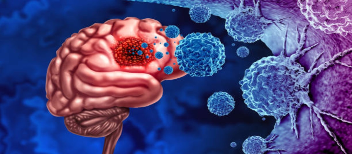
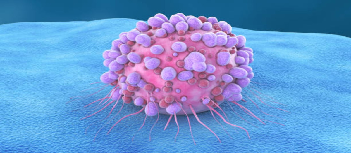


Image Guided Biopsy
Image-guided biopsy is a procedure that uses medical imaging techniques, such as ultrasound, computed tomography (CT), magnetic resonance imaging (MRI), or fluoroscopy, to guide the placement of a biopsy needle to obtain tissue samples from suspicious areas in the body. It is performed to diagnose and characterize various conditions, including tumors, infections, and inflammatory diseases.Here are some key points about image-guided biopsy:1. Procedure: Image-guided biopsy involves the use of real-time imaging to precisely locate and target the suspicious area. The procedure is typically performed by a radiologist or an interventional radiologist. 2. Needle placement: Once the suspicious area is identified on the image, the radiologist uses the guidance of the imaging modality to accurately guide the biopsy needle to the desired location. 3. Tissue sample collection: Once the needle is in the correct position, the radiologist may use various techniques to obtain the tissue sample. This can include using a spring-loaded mechanism to quickly deploy the biopsy needle and collect the tissue sample or performing multiple needle insertions to obtain several samples from different angles or locations within the suspicious area.4. Pathological examination: The collected tissue samples are sent to a pathology laboratory, where a pathologist examines them under a microscope and performs various tests to determine the presence of abnormalities, such as cancer or other diseases.5. Advantages: Image-guided biopsy offers several advantages over other biopsy techniques. It allows for the precise targeting of the suspicious area, reducing the risk of sampling errors and increasing the accuracy of the diagnosis.6. Risks and complications: Image-guided biopsy is generally a safe procedure. However, there is a small risk of bleeding, infection, bruising, or damage to surrounding structures.
READ MORE