+918048036023

DRMISHILPARIKH https://www.mskonco.com
Preview

This is your website preview.

Currently it only shows your basic business info. Start adding relevant business details such as description, images and products or services to gain your customers attention by using Boost 360 android app / iOS App / web portal.

919820800648

Treatments

Image-guided biopsy is a procedure that uses medical imaging techniques, such as ultrasound, computed tomography (CT), magnetic resonance imaging (MRI), or fluoroscopy, to guide the placement of a biopsy needle to obtain tissue samples from suspicious areas in the body. It is performed to diagnose and characterize various conditions, including tumors, infections, and inflammatory diseases.Here are some key points about image-guided biopsy:1. Procedure: Image-guided biopsy involves the use of real-time imaging to precisely locate and target the suspicious area. The procedure is typically performed by a radiologist or an interventional radiologist. 2. Needle placement: Once the suspicious area is identified on the image, the radiologist uses the guidance of the imaging modality to accurately guide the biopsy needle to the desired location. 3. Tissue sample collection: Once the needle is in the correct position, the radiologist may use various techniques to obtain the tissue sample. This can include using a spring-loaded mechanism to quickly deploy the biopsy needle and collect the tissue sample or performing multiple needle insertions to obtain several samples from different angles or locations within the suspicious area.4. Pathological examination: The collected tissue samples are sent to a pathology laboratory, where a pathologist examines them under a microscope and performs various tests to determine the presence of abnormalities, such as cancer or other diseases.5. Advantages: Image-guided biopsy offers several advantages over other biopsy techniques. It allows for the precise targeting of the suspicious area, reducing the risk of sampling errors and increasing the accuracy of the diagnosis.6. Risks and complications: Image-guided biopsy is generally a safe procedure. However, there is a small risk of bleeding, infection, bruising, or damage to surrounding structures.

Sir H. N. Reliance Foundation Hospital and Research Centre, Tower 3rd Floor, Prarthana Samaj, Raja Ram Mohan Roy Rd, Girgaon,

Core needle biopsy is a minimally invasive diagnostic procedure used to obtain tissue samples from suspicious areas in the body for further examination.Here are some key points about core needle biopsy:1. Procedure: During a core needle biopsy, a radiologist, pathologist, or surgeon uses a hollow needle to extract a small cylindrical sample, or core, of tissue from the suspicious area. The procedure is usually performed under local anesthesia, and imaging guidance, such as ultrasound, mammography, or computed tomography (CT), may be used to precisely target the area of interest.2. Sample collection: The needle is inserted into the target area through a small incision or directly through the skin. The physician carefully advances the needle to the suspicious tissue and then applies suction or manually moves the needle back and forth to collect the tissue sample.3. Pathological examination: The collected tissue samples are sent to a pathology laboratory, where they are analyzed by a pathologist.4. Advantages: Core needle biopsy offers several advantages over more invasive procedures, such as surgical biopsy. It is a relatively quick and safe procedure that can be performed on an outpatient basis.5. Limitations: Although core needle biopsy is highly accurate in many cases, there is a small risk of sampling error or false-negative results, meaning that the biopsy may miss the presence of cancer or other abnormalities.6. Risks and complications: Core needle biopsy is generally a safe procedure. However, there is a small risk of bleeding, infection, bruising, or damage to surrounding structures. These risks are relatively low and are usually outweighed by the benefits of obtaining a tissue sample for accurate diagnosis and treatment planning.Core needle biopsy is an important diagnostic tool in modern medicine, allowing for accurate evaluation of suspicious lesions with minimal invasiveness.

Sir H. N. Reliance Foundation Hospital and Research Centre, Tower 3rd Floor, Prarthana Samaj, Raja Ram Mohan Roy Rd, Girgaon,

3D printing and computer navigation technologies have revolutionized the field of cancer surgery, particularly in complex cases that require precise planning and execution.1. 3D printing: 3D printing allows the creation of patient-specific anatomical models based on medical imaging data, such as CT scans or MRI scans. Surgeons can use these models to better understand the patient's unique anatomy, plan the surgery in detail, and simulate complex procedures.2. Surgical planning: By using 3D printed models, surgeons can preoperatively analyze the tumor location, size, and relationship to adjacent structures. This enables better surgical planning, such as determining the optimal approach and identifying potential challenges before entering the operating room.3. Customized implants: 3D printing technology allows for the creation of custom-designed implants or prostheses tailored to the patient's anatomy. In cancer surgery, this can be particularly useful when removing bone or tissue and reconstructing the affected area.4. Computer navigation: Computer navigation systems use preoperative imaging data and real-time tracking to guide surgeons during the procedure. 5. Intraoperative guidance: With computer navigation, surgeons can track their surgical instruments in real-time and ensure accurate resection or removal of the tumor. 6. Minimally invasive surgery: 3D printing and computer navigation technologies also support the advancement of minimally invasive surgical techniques. Surgeons can use preoperative imaging to plan minimally invasive approaches, and computer navigation provides real-time guidance during the procedure.The combination of 3D printing and computer navigation technologies has significantly advanced the field of cancer surgery. These tools enable surgeons to plan and execute procedures with enhanced precision, improve patient outcomes, and promote personalized care.

Sir H. N. Reliance Foundation Hospital and Research Centre, Tower 3rd Floor, Prarthana Samaj, Raja Ram Mohan Roy Rd, Girgaon,

Meet Our Doctor

DR MISHIL PARIKH

DR MISHIL PARIKH

Sir H. N. Reliance Foundation Hospital and Research Centre, Tower 3rd Floor, Prarthana Samaj, Raja Ram Mohan Roy Rd, Girgaon,
Specializes in:
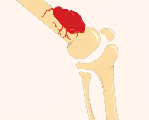
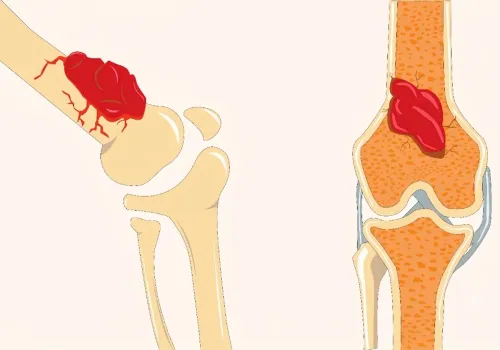
ORTHOPAEDIC ONCOSURGERY
Experience:10 Years
Education:MBBS, MS, MCH, FELLOWSHIP
Mobile:+91-9820800648
Memberships:BOS, IMSOS, ILSOS
Registration No:2011030450

Testimonials

I would like to thank Dr.Mishil Parikh Sir for his assuring support, m here to share my situation from what I was suffering through. I was very afraid of my situation where as I am by profession myself a nurse. I was very panicked but his moral support and encouragement gave me a strength. Thank u so much for the successful treatment of mine with ur excellent support to me and my family. I really want to say that u are such a excellent and brilliant doctor and a good friend which we need in su

*****

I had cancer called Ewing sarcoma on my right leg. The service I received from sir is excellent. His behaviour with patients is so nice and friendly

*****

Latest Update

Bone Cancer Specialist in Mumbai – Expert Orthopedic Oncology Care for Better Outcomes

It's Done!!

Your appointment is confirmed

Doctor Name:

Date & Time:

Contact:

Address: Sir H. N. Reliance Foundation Hospital and Research Centre, Tower 3rd Floor, Prarthana Samaj, Raja Ram Mohan Roy Rd, Girgaon,

Appointment details has also been shared with you on your mobile number . Please arrive atleast ten minnutes ahead of the scheduled time.

treatments