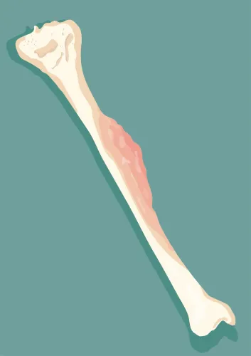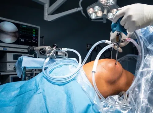

A bone lesion can look alarming on a scan yet be benign, while an early malignant tumor can cause little pain and resemble a less serious problem. You will learn which clues affect urgency, how doctors investigate a lesion, and why imaging, pathology, age, location, and tumor behavior must be considered together.
Key takeaways
- Benign tumors do not spread to distant organs; malignant tumors can metastasize.
- Symptoms, age, and tumor location cannot confirm whether a growth is cancerous.
- Imaging guides evaluation, but a planned biopsy may be needed for diagnosis.
- Treatment ranges from observation to surgery, based on tumor type and behavior.
What benign and malignant bone tumors mean in practice
A benign bone tumor does not spread to distant organs, while a malignant bone tumor can invade nearby tissue and metastasize. That difference changes urgency: suspected malignancy needs prompt imaging, specialist review, and a carefully planned biopsy, while a stable benign lesion may only need observation.
| Type | What it means | Why care differs |
|---|---|---|
| Benign bone tumor | Usually stays in its original site but may enlarge, weaken bone, deform it, or affect a nerve or joint | Observation is reasonable when it is stable; surgery may be needed for pain, growth, fracture risk, or structural damage |
| Malignant bone tumor | Can destroy bone, invade surrounding tissue, and spread through the bloodstream | Delayed diagnosis can allow local invasion or metastasis, so assessment and treatment planning move quickly |
| Locally aggressive benign tumor | Behaves destructively without fitting the usual definition of cancer | Giant cell tumor of bone can enter a joint, recur, and rarely spread to the lungs, so “benign” does not mean harmless |
Pain does not identify the category. Bone tumor symptoms can include persistent pain, bone pain at night, swelling, a lump, reduced joint movement, limping, or a fracture after minor trauma; malignant tumors can initially cause little pain, while benign lesions can hurt when they weaken bone.
Many lesions found incidentally on X-ray, CT, or MRI are not cancer. Worsening pain, pain at rest or at night, a growing mass, unexplained weight loss, fever, or an unexplained fracture deserves prompt medical assessment. A nondiagnostic needle biopsy does not prove benign disease; repeat image-guided or appropriately planned open biopsy may be necessary.
Why symptoms, age, and location cannot identify a tumor alone
Persistent bone pain, swelling or a palpable lump, pain at rest or at night, and a pathological fracture after minor trauma deserve prompt medical assessment. None proves cancer: a benign lesion can hurt by weakening bone, while a malignant growth can begin with little or no pain.
Act sooner if pain is worsening, a lump is enlarging, movement at a nearby joint is becoming limited, or you develop limping, unexplained weight loss, or fever. A fracture through diseased bone needs urgent care because the underlying lesion may require stabilization as well as diagnosis.
Age and skeletal location change the list of possible diagnoses, but they cannot identify one alone. The pattern for a bone tumor in adolescents and adults differs because tumor types have different age and site preferences.
| Age or location | Diagnoses that become more relevant | What the clue cannot establish |
|---|---|---|
| Adolescents and young adults; metaphysis near the knee or upper arm | Osteosarcoma, along with benign lesions and other conditions | A typical site does not confirm osteosarcoma |
| Adults; pelvis, shoulder, or upper limbs | Chondrosarcoma; benign cartilage tumors also occur | Pain or location cannot separate benign from malignant cartilage growth |
| Adults; destructive lesion in any major bone | Metastatic cancer or multiple myeloma, as well as primary bone tumors | The appearance cannot reveal the source without medical evaluation |
Imaging and, when needed, tissue pathology resolve the uncertainty. Do not wait for severe pain before seeking assessment.
How doctors investigate a suspicious or incidental bone lesion
After a lesion appears, doctors do not jump straight to biopsy. They review the images alongside your age, symptoms, prior cancer history, and blood tests. A plain radiograph often shows whether bone has a narrow or wide zone of transition, a well-defined or ill-defined edge, cortical thinning or destruction, and a periosteal reaction.
These patterns indicate how quickly the lesion may be growing, but they do not name it with certainty.
A CT scan bone lesion review clarifies cortical damage, mineralized tumor matrix, small areas of bone formation, and the extent of a fracture. An MRI for bone tumor assessment shows marrow involvement, soft-tissue extension, and the tumor’s relationship to nerves and blood vessels.
For an incidental lesion, doctors commonly follow this sequence:
- Classify the imaging appearance and clinical context. A harmless-looking, stable lesion may need no further work-up; an indeterminate one may need repeat imaging or specialist review.
- Compare with older scans. Stability over time supports a slower process, while growth, cortical destruction, a soft-tissue mass, or aggressive periosteal reaction raises concern.
- Choose biopsy or further imaging when cancer remains possible. A biopsy can confirm the bone tumor diagnosis, but imaging cannot replace pathology when malignancy is suspected.
A needle biopsy must sample the right area and follow the planned surgical route. A nondiagnostic result does not prove benign disease; repeat image-guided biopsy or a carefully planned open biopsy may be necessary.
The conditions that can look alike on imaging
A similar-looking lesion can have a very different risk. Imaging narrows the possibilities, but biopsy or other pathology is needed when cancer remains a serious concern.
| Lesion | Typical imaging clues | Key difference |
|---|---|---|
| Bone cyst | A fluid-filled, sharply outlined cavity, often in a child or adolescent | Usually benign, but thinning can cause a fracture |
| Fibrous dysplasia | “Ground-glass” bone with blending edges | Benign replacement of normal bone; it can weaken or deform bone |
| Osteochondroma | A bony projection whose cortex and marrow continue with the parent bone | Usually benign; a growing cartilage cap after skeletal maturity needs review |
| Enchondroma | A cartilage lesion with rings, arcs, or stippled mineralisation | Enchondroma imaging and diagnosis must distinguish a harmless lesion from cartilage cancer |
| Giant cell tumor | Expansile, lytic lesion reaching the joint surface | Benign but locally aggressive; it can recur and rarely spread to the lungs |
| Osteoid osteoma | A tiny CT-visible nidus surrounded by dense reactive bone | Severe night pain relieved by anti-inflammatory medicine is a useful clue |
| Osteosarcoma | Destruction plus osteoid mineralisation, cortical breach, or a soft-tissue mass | Malignant; chemotherapy and surgery are commonly combined |
| Ewing sarcoma | Permeative destruction, layered periosteal reaction, and a soft-tissue mass | Malignant; systemic chemotherapy is central to treatment |
| Metastatic disease | Multiple lytic or sclerotic lesions, often in an adult | Cancer has spread from another organ |
| Myeloma | Multiple “punched-out” lytic lesions | A plasma-cell cancer, not a primary bone sarcoma |
Age, location, pain, blood tests, MRI, and CT refine the interpretation. A bone cyst or fibrous dysplasia can look alarming after a fracture, while a small malignant lesion can look deceptively contained. Samples must be planned by the treating tumor team.
How the diagnosis determines treatment and specialist care
A low-risk lesion that is painless, stable, and characteristic on imaging may need observation rather than immediate biopsy. A growing, destructive, painful, or structurally weakening lesion needs specialist review, additional imaging, or a planned biopsy; an unplanned biopsy can complicate later surgery.
| Option | What it means | When it applies |
|---|---|---|
| Observation | Repeat examination or imaging at a defined interval | Incidental lesions with reassuring imaging and no concerning symptoms |
| Surgery | Remove the lesion, aiming for a safe margin | Symptomatic benign tumors, unstable bone, or localized tumors that can be completely removed |
| Chemotherapy | Treat cancer cells throughout the body | Osteosarcoma chemotherapy and surgery usually work together; Ewing sarcoma treatment generally combines systemic chemotherapy with surgery and/or radiation |
| Radiation | Deliver focused treatment to the tumor area | Ewing sarcoma, tumors that cannot be safely removed, or selected residual or recurrent disease |
| Reconstruction | Restore strength, length, or joint function after removal | Defects requiring a prosthesis, bone graft, or another structural technique |
| Further referral | Move care to a sarcoma or musculoskeletal oncology team | Uncertain pathology, suspected malignancy, difficult pelvic or spinal location, major blood-vessel or nerve involvement, or possible metastases |
“Benign” does not always mean harmless. Giant cell tumor treatment may require surgery because the tumor can erode bone, enter a joint, recur, or rarely reach the lungs. Dr Mishil Parikh evaluates these treatment decisions within specialist musculoskeletal tumor care, where pathology, imaging, and surgical planning are reviewed together.
Related service
Sarcoma / Musculoskeletal / Orthopaedic Oncology Bone Sarcoma, Benign Bone Tumor Bone sarcoma and benign bone tumors are two distinct types of bone conditions. 1. View service → |
Frequently asked questions
What is the difference between a benign and malignant bone tumor?
A benign bone tumor does not spread to distant organs. A malignant bone tumor can invade nearby tissue and metastasize.
Can symptoms, age, or tumor location identify a bone tumor?
No. Pain, swelling, age, and location can suggest possibilities but cannot determine whether a lesion is benign or malignant alone.
How do doctors investigate a suspicious bone lesion?
Doctors use the clinical history, physical examination, X-rays, MRI, CT or other imaging, and a carefully planned biopsy when tissue diagnosis is needed.
What conditions can look like a bone tumor on imaging?
Infection, healing bone injury, noncancerous lesions, and other bone conditions can resemble a tumor, so imaging findings require clinical and specialist review.
How does diagnosis affect bone tumor treatment?
A stable benign lesion may need observation, while an aggressive or malignant tumor may require specialist surgery, oncology care, and treatment planned around its location and spread.