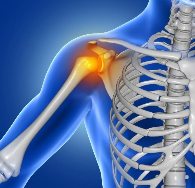
Persistent deep bone pain, swelling, or a growing lump deserves assessment when it does not behave like an ordinary strain or arthritis. By the end, you will know how doctors distinguish suspicious cartilage tumors, why biopsy planning matters, and how subtype, grade, location, and spread shape treatment.
Key takeaways
- Tumor location affects symptoms, biopsy planning, surgical access, and reconstruction.
- Persistent deep pain or a growing lump needs specialist assessment.
- A planned biopsy confirms diagnosis after imaging identifies the safest biopsy path.
- Grade, subtype, margins, and lung spread guide treatment and follow-up.
What chondrosarcoma is and why its location matters
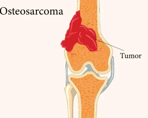
Chondrosarcoma is a malignant cartilage-producing tumor: its cells make abnormal cartilage matrix inside bone or, less often, soft tissue. That distinguishes it from osteosarcoma, which produces malignant osteoid, or immature bone. Chondrosarcoma is not automatically slow or low-risk; grade, size, margin status, local recurrence, and spread to the lungs shape its outlook.
Its subtype changes the level and type of concern:
- Conventional chondrosarcoma is the commonest form. Low-grade tumors can grow slowly, while higher-grade tumors behave more aggressively.
- Clear-cell chondrosarcoma often affects the ends of long bones and can recur or spread despite its relatively bland appearance.
- Mesenchymal chondrosarcoma contains small round cells rather than typical cartilage and can progress aggressively.
- Dedifferentiated chondrosarcoma combines cartilage cancer with a separate high-grade component and generally has a substantially worse outlook than conventional low-grade disease.
Location matters because surgery must remove the tumor without sacrificing critical function. Lesions in the pelvis, hip, shoulder, ribs, or spine sit near nerves, blood vessels, organs, or major joints; local recurrence is harder to control there.
A tumor in an accessible long bone may offer more reconstructive options, while a pelvic or spinal lesion often requires a more complex plan. Even a low-grade tumor therefore deserves location-specific assessment rather than automatic reassurance.
Symptoms that should lead to specialist assessment
Pain that persists, deepens, or continues at rest deserves assessment rather than repeated treatment for a strain or arthritis. Persistent deep pain that lasts several weeks, worsens over time, or wakes you at night is more concerning than soreness that steadily improves after an injury.
A cartilage lesion also needs review when you notice a swelling or growing lump, particularly if it feels fixed, firm, or lies over a bone. A mass deep in the pelvis, shoulder, ribs, or thigh can remain hidden until it becomes sizeable, so the absence of an obvious lump does not exclude a serious lesion.
| Symptom pattern | Why it needs investigation | What to do |
|---|---|---|
| Persistent deep pain, especially at rest or night | A tumor can cause pain without a clear injury | Arrange assessment with an orthopedic or musculoskeletal tumor specialist |
| Swelling or a growing lump | Expansion can affect bone, muscle, or nearby nerves | Seek review promptly rather than monitoring it indefinitely |
| Reduced joint movement | A lesion near the hip, shoulder, or another joint may be restricting movement | Request examination and imaging |
| Sudden severe pain after minor force | A weakened bone can fracture through a lesion | Seek urgent medical care |
Seek urgent attention for sudden inability to bear weight, a new deformity, numbness, or weakness. A prior diagnosis of enchondroma should also be reconsidered if pain, growth, or cortical change develops.
How imaging and a planned biopsy establish the diagnosis
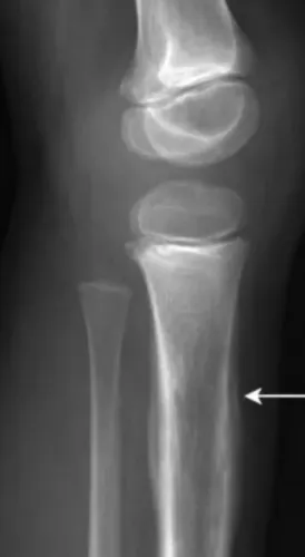
A plain X-ray is usually the first test. Deep endosteal scalloping, cortical thinning or destruction, a soft-tissue mass, and rings-and-arcs or stippled mineralization raise concern. The X-ray cannot show the full tumor or confirm malignancy.
| Test | What it defines | Important limitation |
|---|---|---|
| Plain X-ray | Bone destruction, chondroid mineralization, and fracture risk | Does not show full local extent |
| CT scan | Mineralized cartilage, cortical breach, and subtle bone destruction | Does not replace tissue diagnosis |
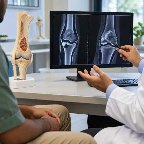
| MRI with contrast | Marrow involvement, soft-tissue extension, joint and neurovascular relationships, and the safest biopsy route | Imaging appearance alone cannot confirm cancer |
| Chest CT staging | Small lung metastases, the commonest site of distant spread | Does not diagnose the primary bone lesion |
A planned image-guided core-needle biopsy confirms whether the lesion is chondrosarcoma and identifies its subtype and grade. The biopsy track must be placed where it can be removed during definitive surgery; an improvised route can contaminate otherwise usable tissue and complicate limb-sparing treatment.
Sampling matters because cartilage tumors are heterogeneous. A small specimen can undergrade a lesion, especially when imaging shows areas with different levels of aggression or possible dedifferentiation, so the grade can change after the resection specimen is examined. Distinguishing low-grade chondrosarcoma from enchondroma requires pathology alongside pain, growth, size, location, cortical involvement, and imaging.
IDH1 or IDH2 mutations can support classification, but neither mutation alone proves the diagnosis or grade.
How grade and subtype change the treatment decision
A biopsy result does not determine treatment by itself. Pathologists combine the tumor grade and subtype with imaging, pain, growth, cortical destruction, and location—especially when distinguishing low-grade chondrosarcoma from an enchondroma is difficult on a small sample.
| Subtype | Typical treatment implication | Why |
|---|---|---|
| Conventional chondrosarcoma | Wide en-bloc surgery | Low-grade lesions may be less aggressive, but conventional tumors respond poorly to standard chemotherapy and photon radiotherapy. |
| Clear-cell chondrosarcoma | Surgery with careful long-term surveillance | It can recur or spread despite a deceptively lower-grade appearance. |
| Mesenchymal chondrosarcoma | Surgery plus consideration of chemotherapy | This subtype behaves aggressively and is more chemotherapy-sensitive than conventional disease. |
| Dedifferentiated chondrosarcoma | Surgery plus oncology planning for systemic treatment | A high-grade dedifferentiated component carries a substantially higher risk of metastasis. |
For a resectable conventional tumor, wide surgery with a planned margin is usually the curative treatment. Pelvic, spinal, or hip tumors demand different operations from lesions in an accessible long bone; nearby nerves, major vessels, joints, and organs may limit the margin or require reconstruction. Limb salvage does not guarantee normal function.
Chemotherapy is not routine for most conventional chondrosarcomas. The team considers it more strongly for mesenchymal chondrosarcoma and selected dedifferentiated tumors, alongside grade, size, metastases, and the patient’s fitness.
Radiotherapy becomes useful when surgery cannot safely remove the tumor, a margin is positive or very close and further surgery is not feasible, the tumor recurs, or pain needs control.
A specialist such as Dr Mishil Parikh helps align the pathology review with the surgical anatomy before committing to an operation. An IDH1 or IDH2 mutation can support classification, but it does not establish grade or automatically create a targeted-treatment option.
Related service
Sarcoma / Musculoskeletal / Orthopaedic Oncology Bone Sarcoma, Benign Bone Tumor Bone sarcoma and benign bone tumors are two distinct types of bone conditions. 1. View service → |
Surgery, margins, reconstruction, and follow-up
Once the plan is chosen, wide en-bloc surgery removes the tumor in one piece with a surrounding cuff of healthy tissue. The aim is negative margins: no cancer cells at the cut edge. If the tumor lies near a major nerve, vessel, joint, or pelvic structure, achieving that margin can make the operation technically demanding.
Reconstruction restores stability and movement after bone or soft tissue is removed. It may use a bone graft, metal implant, joint replacement, or pelvic reconstruction. Limb salvage is often possible, but keeping the limb does not guarantee normal strength, sensation, walking, or joint movement.
Recovery may include:
- Wound care and pain control
- Physiotherapy to regain movement, balance, and strength
- Protected weight-bearing until the reconstruction heals
- Occupational support for daily activities, work, or mobility aids
A pathology report confirming negative margins does not end care. Local recurrence can develop at the original site, and lung metastases can appear after apparently complete removal, sometimes years later. Follow-up is adjusted to the tumor’s grade, subtype, location, margin status, and time since surgery.
Surveillance usually combines:
- Examination of the surgical site and overall function
- Periodic MRI, CT, or other imaging of the original tumor site
- Chest imaging to check for spread to the lungs
Report new or increasing pain, swelling, a lump, reduced movement, or breathing symptoms between scheduled visits. Detecting recurrence early can preserve more treatment and function.
Frequently asked questions
What is chondrosarcoma, and why does its location matter?
Chondrosarcoma is a malignant cartilage-producing tumor. Its location affects symptoms, biopsy planning, surgical access, nearby structures, and reconstruction.
Which symptoms of chondrosarcoma need specialist assessment?
Persistent deep pain, pain that worsens at night, swelling, a growing lump, reduced movement, or unexplained fractures should lead to specialist assessment.
How is chondrosarcoma diagnosed?
Imaging defines the tumor’s size and location, while a planned biopsy examines tissue and confirms the diagnosis. The biopsy route must be chosen with the definitive surgery in mind.
How do grade and subtype affect chondrosarcoma treatment?
Grade and subtype indicate tumor behavior and help determine whether surgery alone is appropriate or whether additional treatment and closer surveillance are needed.
How is chondrosarcoma treated after diagnosis?
Surgery removes the tumor with planned margins. The surgical team then considers reconstruction, margin results, local recurrence, lung spread, and long-term follow-up.