Persistent bone pain, swelling, limping, or a fracture after minor trauma deserves a clearer explanation than “just a sports injury” or growing pains. By the end, you will know which symptoms need assessment, how doctors confirm osteosarcoma, why biopsy planning matters, and how chemotherapy, surgery, radiation, and follow-up fit together.
Key takeaways
- Persistent bone pain with swelling needs prompt medical assessment.
- MRI shows the local tumor; biopsy confirms whether it is osteosarcoma.
- Treatment commonly combines chemotherapy with limb-sparing surgery.
- Regular imaging follow-up checks for recurrence and lung spread.
What Osteosarcoma Looks Like and Who Develops It
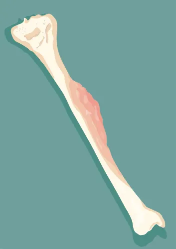
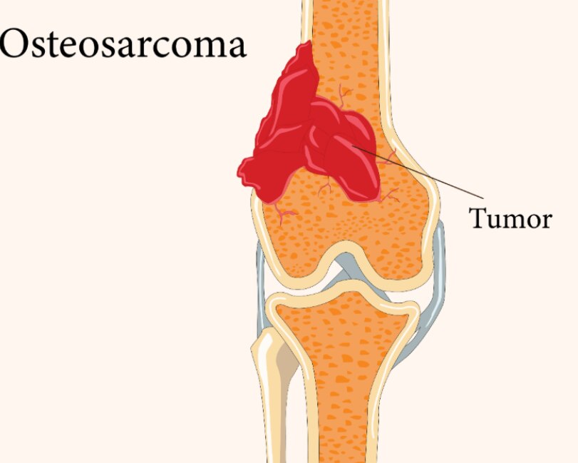
Persistent, localized bone pain that worsens or occurs without a clear injury deserves assessment, especially with swelling. Osteosarcoma begins in osteoblast-producing bone-forming cells. Its defining feature is malignant cells producing osteoid, or immature bone, which distinguishes it from other bone sarcomas.
It most often appears during an adolescent growth spurt, but a separate group of older adults also develops it. Common sites include the lower femur and upper tibia around the knee, and the upper arm near the shoulder. The pelvis and other long bones are possible sites; location alone does not confirm or exclude osteosarcoma.
Possible osteosarcoma symptoms include:
- Persistent bone pain at night or after activity
- Swelling or a palpable lump
- Reduced movement at a nearby joint
- Limping
- A fracture after minor trauma
| Pattern | More reassuring explanation | Less reassuring pattern |
|---|---|---|
| Sports injury | Pain follows a clear impact and steadily improves | Pain persists, progresses, or starts without an injury |
| Growing pains | Intermittent aching without swelling or a fixed tender spot | Localized pain with swelling, a lump, or worsening symptoms |
| Arthritis | Joint-centered stiffness and pain, usually in an older adult | Deep bone pain with a growing mass or reduced movement |
| Benign bone cyst | Often found incidentally or after a fracture | Progressive pain, swelling, or fracture after minor trauma |
These signs do not diagnose cancer, but persistence, progression, or swelling makes an examination important.
When Symptoms Need Urgent Assessment and What Happens First
Sudden severe pain with inability to bear weight or use a limb needs urgent assessment, especially after minor trauma. Rapidly enlarging swelling also requires prompt review because it can indicate an aggressive bone process.
Seek urgent care if you have:
- Severe, sudden pain with inability to walk, stand, or use the affected arm
- Swelling that enlarges quickly or becomes tense and markedly painful
- A suspected pathological fracture, meaning a break through bone weakened by an underlying lesion
The first medical evaluation usually includes a physical examination, assessment of movement and nerve or blood-vessel function, and a plain X-ray. Blood tests are added when indicated; they can assess general health and abnormalities such as raised alkaline phosphatase, but they cannot confirm osteosarcoma.
An osteosarcoma X-ray may show a destructive bone lesion, mixed lytic and sclerotic change, cortical destruction, or aggressive periosteal reaction. A sunburst pattern or Codman triangle is a warning sign, not proof of cancer.
If imaging raises concern, your clinician should refer you to an orthopedic oncology or musculoskeletal tumor team before biopsy or definitive treatment. An X-ray can raise suspicion but cannot confirm osteosarcoma. Location or appearance alone does not establish the diagnosis, because infections, benign tumors, and other bone cancers can look similar.
How Doctors Confirm and Stage Osteosarcoma
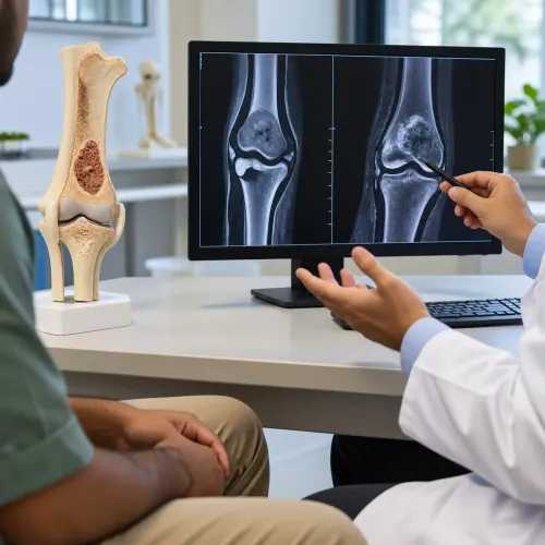
MRI of the entire involved bone and adjacent joint maps marrow involvement, soft-tissue extension, and skip lesions—separate tumor foci in the same bone or nearby region. These findings can change the planned surgical margins.
| Test | What it shows | Why it matters |
|---|---|---|
| MRI | Marrow, soft-tissue spread, and skip lesions | Defines the local operation |
| Chest CT | Pulmonary metastases | More sensitive than chest X-ray for lung spread |
| Whole-body bone imaging | Other skeletal lesions | Detects possible bone metastases |
| PET/CT | Metabolic tumor activity and distant disease | Used when clinically appropriate |
A biopsy is required for an osteosarcoma diagnosis. The orthopedic oncology team performing definitive surgery must handle biopsy tract planning: an incorrectly placed tract can contaminate additional tissue compartments, enlarge the required resection, or compromise limb-salvage surgery. Surgeons usually remove the biopsy tract with the tumor.
A specialist pathologist experienced in bone and soft-tissue tumors reviews the specimen. The hallmark of conventional osteosarcoma is malignant tumor cells producing osteoid, or immature bone, directly. Imaging and pathology must be interpreted together because several bone lesions can mimic osteosarcoma, and a biopsy result without its imaging context can misclassify the disease.
Staging determines whether surgery can remove all visible disease, whether lung or other metastases need treatment, and how chemotherapy is planned. A chest CT, whole-body imaging, and the MRI findings together provide the map needed to choose the safest operation and the appropriate treatment sequence.
Which Osteosarcoma Treatments Are Used and Why
Most resectable, high-grade conventional osteosarcomas need osteosarcoma treatment with chemotherapy before and after surgery, not surgery alone. The common backbone is MAP chemotherapy: high-dose methotrexate, doxorubicin, and cisplatin. Doctors adjust the regimen for age, fitness, relapse risk, organ function, and local practice.
| Option | Why it is used | Key considerations |
|---|---|---|
| Chemotherapy | Treats microscopic disease throughout the body | Monitor doxorubicin with cardiac-function tests; cisplatin can injure kidneys and hearing; high-dose methotrexate requires drug-level and kidney monitoring |
| Surgery | Removes the primary tumour with adequate margins | Reconstruction may use a metallic endoprosthesis, allograft, or another method |
| Radiation therapy | Treats selected unresectable, incompletely resected, or recurrent tumours | Not routine when complete removal is feasible because osteosarcoma responds less reliably than many other cancers |
| Metastatic treatment | Controls disease outside the primary site | Selected patients with lung-only metastases may undergo lung metastasectomy if all detectable deposits can be removed, often with chemotherapy |
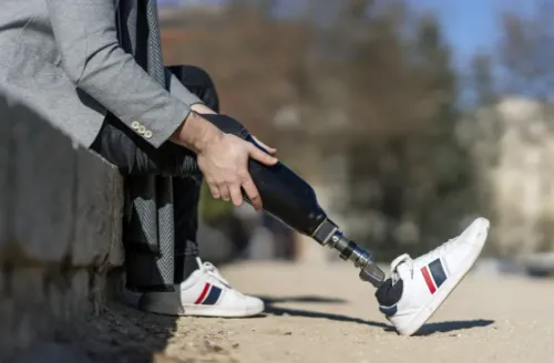
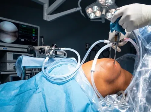
Limb-sparing surgery is suitable when surgeons can remove the tumour safely and leave a functional limb. Amputation is appropriate when major nerves, major vessels, or extensive tissue make a safe, functional resection impossible.
Treatment planning should include fertility preservation before chemotherapy when relevant. Doctors also weigh tumour location, margins, metastases, and expected function rather than choosing one treatment in isolation.
What Treatment Response, Recovery, and Follow-Up Mean
About 90% or more tumor necrosis percentage in the surgical specimen after preoperative chemotherapy is generally considered a good histologic response. Less necrosis is associated with a higher relapse risk, but neither result guarantees success or failure; it is one part of your osteosarcoma prognosis.
Ask the treatment team:
- What did the pathology report show, including the necrosis percentage, margins, tumor grade, and response to chemotherapy?
- What movement, strength, gait, and daily activities should reconstruction or amputation allow?
- What rehabilitation will I need, and when can physiotherapy begin?
- Which scans and examinations will track the original tumor site and lungs, and how often will osteosarcoma follow-up occur?
Rehabilitation focuses on restoring joint movement, muscle strength, balance, gait, and safe daily function. Recovery after limb reconstruction or amputation takes time, and the plan may include physiotherapy, occupational therapy, gait training, prosthetic fitting, pain control, and adjustments as your function improves.
Surveillance examines the original site because local recurrence can occur, and checks the chest because osteosarcoma can recur in the lungs. Your treating team sets the schedule according to the tumor, surgery, treatment response, and overall risk.
A suspected bone lesion deserves an orthopedic oncology or musculoskeletal tumor service for coordinated imaging review, biopsy planning, surgery, reconstruction, and long-term care. Dr Mishil Parikh in Mumbai is one clinician patients may consider for this coordinated evaluation. Referral does not prove cancer; it protects diagnostic and treatment options while osteosarcoma remains a possibility.
Related services
Sarcoma / Musculoskeletal / Orthopaedic Oncology Bone Sarcoma, Benign Bone Tumor Bone sarcoma and benign bone tumors are two distinct types of bone conditions. 1. View service → |
Limb Salvage Surgery Limb salvage surgery, also known as limb-sparing surgery, is a procedure performed to remove a tumor or manage a severe injury in a limb while... View service → |
Frequently asked questions
What are common symptoms of osteosarcoma?
Osteosarcoma often causes persistent localized bone pain, swelling, tenderness, reduced movement, or a lump. Pain that worsens at night or occurs without a clear injury needs assessment.
When does suspected osteosarcoma need urgent assessment?
Arrange prompt assessment for persistent bone pain, swelling, a growing lump, unexplained reduced movement, or a fracture after minor trauma. Severe pain, sudden loss of function, or a suspected fracture requires urgent care.
How do doctors confirm and stage osteosarcoma?
Doctors use examination and imaging such as X-rays, MRI, CT, and chest imaging, then confirm the diagnosis with a planned biopsy. Staging assesses the primary tumor, nearby tissues, and spread, especially to the lungs.
What treatments are used for osteosarcoma?
Treatment commonly combines chemotherapy and surgery. Surgeons may remove the tumor while preserving the limb when safe, followed by reconstruction; amputation is considered when adequate tumor clearance and function cannot be achieved.
What does follow-up after osteosarcoma treatment involve?
Follow-up includes scheduled examinations and imaging of the original tumor site and lungs. The schedule also monitors treatment effects, recovery, reconstruction, physical function, and signs of recurrence.