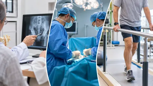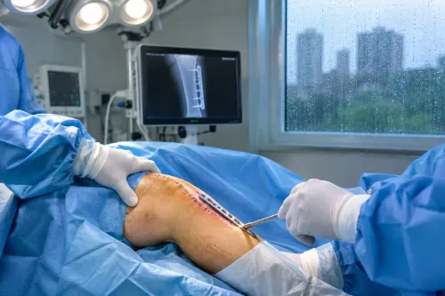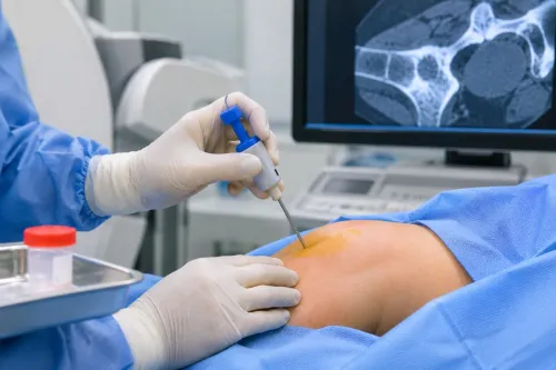
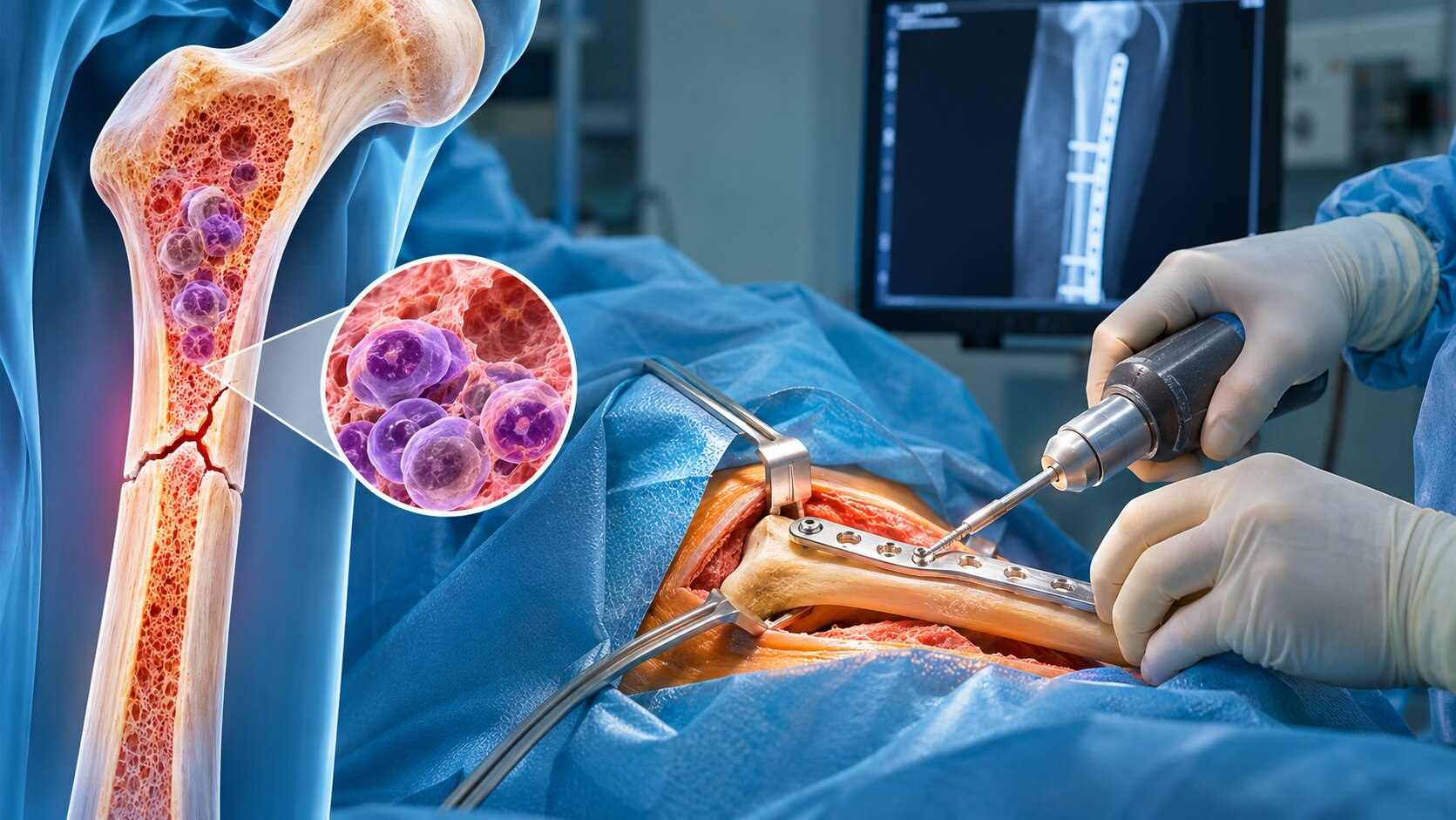
A myeloma bone lesion does not automatically need surgery, but waiting for a break can make treatment more extensive when the bone is already unstable. By matching symptoms, imaging, lesion location and functional limitation to the right risk framework, you can understand when observation, radiotherapy, prophylactic fixation or reconstruction deserves urgent specialist review.
Key takeaways
- Seek same-day assessment after sudden pain, deformity, or inability to bear weight.
- Doctors combine imaging, lesion location, pain, and bone strength to estimate fracture risk.
- Prophylactic fixation can prevent a break when a weakened bone is unstable.
- Ask which operation preserves function and how myeloma treatment affects timing.
Is This an Impending Fracture or an Emergency?
A sudden snap, deformity, or inability to bear weight or use an arm needs same-day emergency assessment because the lesion may already have caused a pathological fracture.
New leg weakness, numbness, loss of bladder or bowel control, or rapidly worsening back pain is also an emergency: spinal cord or nerve compression cannot wait for a routine appointment.
- Same-day emergency care: sudden severe pain after a minor movement or fall, a visibly shortened or crooked limb, inability to walk, or new neurological symptoms.
- Urgent surgical planning: persistent pain when standing, walking, lifting, or turning in bed; a lytic lesion that has removed much of the cortex; a visible fracture line; progressive bone collapse; or a vertebral lesion with posterior-wall damage, epidural extension, or worsening alignment.
- Continued monitoring: little or no pain, preserved function, an intact or well-supported cortex, no fracture line or collapse, and stable findings on comparison imaging.
An impending fracture means the bone has not broken but lacks enough strength for normal loading. X-rays show cortical thinning, a lytic defect, or alignment change; CT defines the remaining shell and fracture geometry, while MRI detects marrow spread, soft-tissue mass, and spinal or nerve involvement.
Radiotherapy can relieve tumour pain but does not reliably rebuild a mechanically unsafe femur or vertebra.
Ask the team to state whether the proposed treatment is observation, radiotherapy, cement augmentation, or prophylactic fixation, and what imaging supports it. When comparing myeloma bone surgery options in Mumbai, choose a service that can coordinate haematology, orthopaedic oncology, radiology, pathology, spine care, radiation oncology, anaesthesia, and rehabilitation.
How Doctors Calculate Fracture Risk Before a Break
Doctors estimate fracture risk from the bone’s structure, your pain and your ability to use the limb—not from tumour size alone. If you are deciding which myeloma for bone surgery option fits in mumbai, request a site-specific review by haematology, orthopaedic oncology or spine surgery, radiology and radiation oncology.
1. For a long-bone lesion, calculate the Mirels score. Score site, pain, lesion type and the proportion of bone diameter involved from 1 to 3 each. A score of 9 or more supports prophylactic stabilisation; 8 requires individual review; 7 or less usually favours observation and myeloma treatment.
A lytic lesion, severe functional pain or extensive cortical loss can shift the decision even when the total is borderline.
2. For the spine, use MRI and CT alongside the Spinal Instability Neoplastic Score. SINS measures location, mechanical pain, lesion quality, alignment, vertebral-body collapse and posterior-element involvement. Scores of 7–12 indicate possible instability, while 13–18 indicate instability and require specialist spine assessment.
3. For the pelvis and hip, doctors examine weight-bearing pain, gait, CT evidence of acetabular or femoral-neck destruction, and pelvic-ring or joint stability. For the shoulder, they assess proximal-humerus cortical loss, pain during lifting and MRI or CT findings.
4. For ribs, imaging checks cortical breach, displacement and chest-wall or pleural complications. Rib lesions rarely need preventive fixation unless instability, severe persistent pain or respiratory complications develop.
When Observation or Prophylactic Fixation Makes Sense
1. Choose observation when the lesion is structurally stable, pain is mild or improving, you can use the limb normally, and imaging shows no major cortical destruction, collapse or displacement. Continue systemic myeloma treatment, prescribed analgesia and activity limits, with repeat imaging at the interval your team sets.
New pain during walking or lifting changes the decision.
2. Choose prophylactic fixation when pain is mechanical, function is declining, or a long-bone lesion has extensive cortical loss, a large lytic defect or visible deformity. Stabilising the bone before it breaks usually preserves more function and avoids the displacement, blood loss and rehabilitation burden of repairing a completed pathological fracture.
Weight-bearing pain deserves prompt reassessment, not a test of endurance.
3. Do not treat severe mechanical weakness with radiotherapy alone. Radiotherapy can reduce tumour-related pain, but it does not reliably restore the strength of a badly destroyed femur, humerus or vertebra. Depending on the site, the safer plan may combine radiotherapy with a nail, plate, cement augmentation, vertebral procedure or joint replacement.
4. Confirm the diagnosis before definitive implants when the lesion is the first sign of disease or scans cannot distinguish myeloma from plasmacytoma, metastasis, infection or a primary bone tumour. Ask the team to coordinate haematology, orthopaedic oncology, radiology, pathology, radiation oncology and rehabilitation.
If you are comparing myeloma fracture surgery in Mumbai, ask whether the proposed operation is prophylactic or treats an established fracture, and what finding would change it.
Choosing the Operation by Bone, Lesion and Stability
The best operation matches the bone’s remaining strength, the lesion’s position and whether the joint or spine has failed. A shaft lesion with usable bone differs greatly from a destroyed hip end or unstable pelvic ring.
| Option | Best fit | Durability, recovery and risk |
|---|---|---|
| Intramedullary nail | Long-bone lesions spreading through the femur or humerus | Protects a long segment and often enables early walking or arm use; risks include blood loss, infection, nail failure and damage to nearby structures. |
| Plate and screws | Short-segment or metaphyseal defects with enough bone for screw purchase | Gives precise support but covers less bone; recovery depends on fracture healing, and screws can loosen in weak bone. |
| Cement augmentation | A large cavity or poor residual bone, often combined with fixation | Polymethylmethacrylate improves immediate stability; leakage, thermal injury, infection and mechanical failure remain possible. |
| Joint replacement | A destroyed joint surface or major bone end, such as the proximal femur | Provides immediate structural support when screws or nails cannot hold; it is a larger operation with dislocation, infection and blood-clot risks. |
| Vertebral augmentation | Painful vertebral collapse without a need for open decompression | Can relieve mechanical pain through cement injection, with shorter recovery; cement leakage and nerve injury are important risks. |
| Pelvic reconstruction | An unstable pelvic ring or acetabular defect affecting weight-bearing | Restores alignment and load transfer but involves demanding surgery, longer rehabilitation and substantial bleeding risk. |
Radiotherapy can reduce tumour pain but cannot reliably rebuild a severely weakened weight-bearing bone. Comparing myeloma bone surgery options in Mumbai therefore requires haematology, orthopaedic oncology or spine, radiology, anaesthesia and rehabilitation input. Cement or surgery also does not replace systemic anti-myeloma treatment.
What to Ask a Myeloma Bone Surgery Team in Mumbai
Ask for the decision in writing: is the operation prophylactic, for an established pathological fracture, or for instability, nerve compression or uncontrolled mechanical pain? A myeloma orthopedic surgeon in Mumbai should show the CT or MRI findings supporting that choice, not rely on an X-ray appearance alone.
| Team evidence | Question to ask | Why it matters |
|---|---|---|
| Routine tumour-fracture work | How many pathological and impending fractures do you treat? | Routine trauma fixation may not address diseased bone. |
| Multidisciplinary review | Will haematology, orthopaedic oncology, radiology, pathology, radiation oncology and rehabilitation review my case? | Surgery must fit systemic treatment and radiotherapy. |
| Diagnostic certainty | Is biopsy needed before implants, and what changes if pathology shows plasmacytoma, metastasis, infection or another tumour? | Fixation can complicate later diagnosis and treatment. |
| Reconstruction capability | If the joint end is destroyed, can you compare fixation with cement, arthroplasty or reconstruction? | Screws or nails fail when too little bone remains. |
Ask which scans measured lesion size, cortical destruction, joint involvement and spinal or pelvic stability. Request the alternative: observation, radiotherapy, cement augmentation or surgery, with the consequence of waiting.
Also ask when chemotherapy, zoledronic acid or denosumab will resume; who checks calcium, vitamin D and kidney function; and what weight-bearing, walking aid, wound review and physiotherapy schedule you will follow. Myeloma bone tumor care in Mumbai should include a named contact for these handoffs.
Whether you consult Dr Mishil Parikh or another team, compare documented reasoning, not a promise of a particular implant.
Frequently asked questions
How can you tell whether a myeloma bone problem is an impending fracture or an emergency?
A sudden snap, deformity, or inability to bear weight or use an arm needs same-day emergency assessment because a pathological fracture may have occurred.
How do doctors calculate fracture risk before a myeloma bone breaks?
They assess pain, imaging findings, the lesion’s size and location, bone destruction, and mechanical stability before choosing observation or fixation.
When does prophylactic fixation make sense for myeloma bone disease?
Fixation is considered when a lesion has weakened a load-bearing bone or creates a high risk of fracture, while stable lesions may be observed with oncology treatment and follow-up.
How does the affected bone change the myeloma surgery option?
The operation depends on the bone involved, lesion position, remaining bone strength, joint involvement, and whether the goal is stabilisation, reconstruction, or fracture treatment.
What should you ask a myeloma bone surgery team in Mumbai?
Ask about fracture risk, the recommended operation, alternatives, functional recovery, surgical timing alongside myeloma treatment, and the team’s experience with musculoskeletal tumour care.
Keywords