Monitoring is reasonable only when a clinician has established that a lump is benign or has classified it as low risk and set a review plan. By the end, you will know which findings require MRI and biopsy, when planned surgery is safer than observation, and what a coordinated Mumbai sarcoma team should provide.
Key takeaways
- Monitor only with a documented diagnosis, review date, measurements and warning symptoms.
- Growing, deep, painful or enlarging lumps need prompt specialist assessment.
- A biopsy route must be planned with the surgeon who may perform the operation.
- Treatment depends on tumor type, size, depth, spread and functional goals.
When Is Monitoring Safe for a Soft-Tissue Lump?
Monitoring is defensible when a clinician has identified a small, superficial, stable lesion and made a confident benign diagnosis. It is not permission to ignore a lump: the written plan should specify the examination interval, repeat ultrasound or MRI date, measurements to record, and symptoms that require earlier review.
A painless or small mass can still be sarcoma.
Observation becomes unsafe when any of these features is present:
- The lump is growing, painful, or fixed to surrounding tissue.
- It measures around 5 cm or more.
- It lies deep beneath the fascia.
- It has returned after previous removal.
- Ultrasound or MRI findings are uncertain or do not support a clear benign diagnosis.
Ask these questions before agreeing to surveillance:
- What diagnosis supports monitoring?
- When will the lump be measured again?
- Will the next test be ultrasound or MRI?
- Who will review the scan?
- What change requires immediate contact?
If you are comparing soft tissue sarcoma surgery or monitoring in Mumbai, seek urgent specialist assessment for a growing, deep, recurrent, large, painful, fixed, or uncertain mass. A defined surveillance plan can avoid unnecessary treatment for a genuinely benign lesion, while specialist review prevents delayed diagnosis when imaging or behaviour raises concern.
Do not substitute periodic scans arranged without a documented diagnosis and review schedule for a proper assessment.
How Doctors Test Whether a Tumor Is Benign or Sarcoma
A reliable diagnosis moves from clinical assessment to imaging and planned tissue sampling; no single scan can prove that a lump is benign or sarcoma. Your clinician records how long it has been present, whether it is growing or painful, and examines its depth, mobility, size and relation to nearby structures.
1. Ultrasound gives an initial look at a superficial lesion. It can show whether the lump is solid or fluid-filled and guide further assessment, but it does not replace MRI when the mass is deep, enlarging or suspicious.
2. MRI with contrast is the preferred local study for a deep or suspicious mass. It maps size, tissue compartments, fascia, muscle, nerves, vessels and possible skip lesions, which helps surgeons plan treatment while preserving function where possible. MRI still cannot establish the tumour’s exact type or grade.
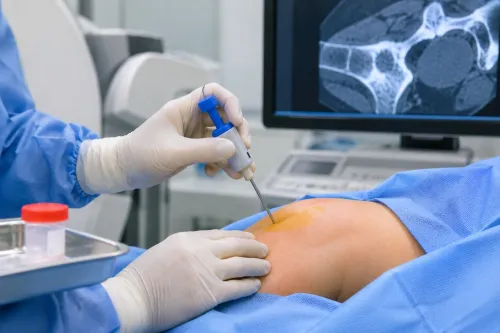
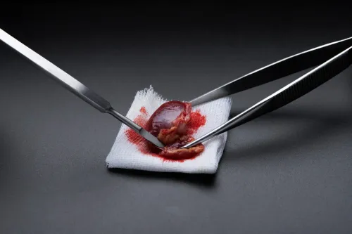
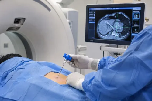
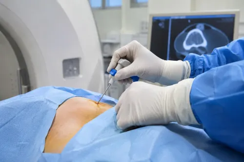
3. A planned core-needle biopsy usually supplies the tissue needed for diagnosis, subtype and grade. The biopsy route must be chosen with later surgery in mind rather than treating it as a separate procedure.
4. Once sarcoma is suspected or confirmed, chest imaging enters staging because the lungs are a frequent site of spread. Abdominal, pelvic, bone or whole-body scans depend on the subtype, tumour location and symptoms; they are not automatic for every patient.
5. Ask for expert sarcoma pathology review when a core report names a rare subtype or uses terms such as “spindle-cell tumour,” “atypical lipomatous tumour” or “sarcoma cannot be excluded.” This review is important when comparing soft tissue options in Mumbai, because an interpretation can change monitoring, surgery and additional treatment.
Why the Biopsy Plan Can Change the Operation
The biopsy choice can determine how much tissue the definitive operation must remove. A suspected sarcoma needs a planned sample, not a quick lump removal.
| Biopsy type | What it does | When it fits |
|---|---|---|
| Core-needle biopsy | Usually supplies tissue for subtype and grade | Preferred for many suspected sarcomas |
| Incisional biopsy | Removes part of the mass | Large or difficult tumours |
| Excisional biopsy | Removes the entire lesion | Selected small, superficial masses after assessment |
Plan the biopsy track along the likely surgical incision, usually longitudinally through a limb. The surgeon can then remove the track with the tumour; a poorly positioned track can contaminate tissue planes and force additional resection. The team performing or coordinating the biopsy should be able to provide definitive treatment.
Do not have a lump removed before imaging and biopsy if sarcoma is possible. Unplanned excision can spread tumour cells into surrounding planes, complicate margin assessment, require wider surgery and reduce function-preserving options. That risk matters when comparing soft tissue tumor surgery in Mumbai.
After an unplanned removal, take these records for a second opinion:
- The complete pathology report
- The original slides or tissue block
- MRI images, not only the written report
- The operative note
- Earlier scans
Ask for specialist review when the report says “spindle-cell tumour,” “atypical lipomatous tumour” or “sarcoma cannot be excluded.” A coordinated review can establish whether further imaging, re-excision or surveillance is appropriate.
What Surgery, Radiation and Chemotherapy Each Contribute
For a confirmed, localized, resectable limb or trunk sarcoma, planned wide excision is usually the main curative treatment. The surgeon removes the tumor with a rim of normal tissue and aims for a negative microscopic margin, meaning no tumor cells reach the examined edge.
| Option | Main contribution | Usual role |
|---|---|---|
| Surgery | Removes the primary tumor | Main treatment for a resectable localized sarcoma |
| Radiation | Reduces local recurrence risk | Added selectively before or after surgery |
| Chemotherapy | Treats susceptible cancer cells throughout the body | Considered for selected high-risk or metastatic disease |
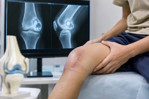
Amputation is not the default. It is considered when complete tumor control and a useful, functional limb cannot be achieved with limb-sparing surgery, reconstruction or other combined treatment.
Radiation is more strongly considered for a deep, large or high-grade tumor, or when the margin is close or uncertain. Preoperative radiation uses a smaller field and lower dose but causes more wound-healing problems; postoperative radiation has more late tissue effects.
Chemotherapy does not routinely replace surgery. Its value depends on the subtype, stage, size, site and your fitness, with greater consideration for selected high-risk localized tumors or metastatic disease than for every small, low-grade lesion.
Follow-up checks for local recurrence and lung metastases on a risk-adapted schedule, using examination, local imaging and chest imaging. For people comparing soft tissue sarcoma options in Mumbai, ask how the team will coordinate surgery, radiation, pathology and medical oncology around your tumor’s specific risks.
How to Choose a Mumbai Team for Surgery or Surveillance
A complete Mumbai pathway should match the tumor’s depth, growth, imaging and diagnosis—not default to surgery. For soft tissue sarcoma surgery or monitoring in Mumbai, check whether one team can coordinate:
- Specialist examination and MRI
- Image-guided core biopsy
- Sarcoma-experienced pathology review
- Chest staging when sarcoma is suspected or confirmed
- Orthopedic-oncology surgery
- Radiation oncology and medical oncology
- Multidisciplinary review
| Pathway | What it means | When it applies |
|---|---|---|
| Planned surgery | Imaging and biopsy guide oncologic margins, reconstruction and function-preserving treatment | A resectable tumor has concerning or confirmed malignant features |
| Structured surveillance | A clinician documents the benign or low-risk diagnosis, examination interval, repeat scan date and triggers for earlier review | A small, superficial, stable lesion has a confident diagnosis |
| Limited clinic pathway | Quick excision or periodic ultrasound without full staging and specialist review | Not enough to establish safe monitoring for a deep, growing or uncertain mass |
Seek a second opinion if the report says “spindle-cell tumor,” “atypical lipomatous tumor,” or “sarcoma cannot be excluded.” Do the same after removal without planned oncologic margins; carry the pathology report, slides or tissue block, MRI images, operative note and earlier scans.
Dr Mishil Parikh, an orthopedic oncology and musculoskeletal cancer surgeon in Mumbai, can help determine whether a mass needs planned surgery, further work-up or coordinated specialist management. A sarcoma specialist in Mumbai—or a soft tissue sarcoma doctor in Mumbai—should be able to explain why surveillance fits your tumor’s risk and anatomy.
Related services
Soft Tissue Tumor A soft tissue tumor refers to an abnormal growth of cells that occurs in the soft tissues of the body. View service → |
Limb Salvage Surgery also known as limb-sparing surgery, is a procedure performed to remove a tumor or manage a severe injury in a limb while preserving the limb itself. View service → |
Frequently asked questions
When is monitoring safe for a soft-tissue lump?
Monitoring is defensible when a clinician has made a confident benign diagnosis for a small, superficial, stable lesion. The plan should include examination intervals, repeat ultrasound or MRI dates, measurements and symptoms requiring earlier review.
How do doctors test whether a tumor is benign or sarcoma?
Doctors combine examination and imaging, such as ultrasound or MRI, with a planned biopsy when the diagnosis remains uncertain. The pathology result identifies the tumor type and helps determine treatment.
Why can the biopsy plan change the operation?
The biopsy track can contain tumor cells, so surgeons plan its location and direction to allow removal during definitive surgery. An unplanned biopsy can complicate later surgery and reconstruction.
What do surgery, radiation and chemotherapy each contribute?
Surgery removes the tumor while aiming to preserve function. Radiation helps control local disease in selected cases, while chemotherapy treats specific sarcoma subtypes or disease that has spread.
How do you choose a Mumbai team for surgery or surveillance?
Ask whether the team regularly treats soft-tissue sarcoma, reviews imaging and pathology together, plans biopsies before surgery, and explains surveillance intervals, treatment alternatives and functional effects.
Related service
Image Guided Biopsy Image-guided biopsy is a procedure that uses medical imaging techniques, such as ultrasound, computed tomography (CT), magnetic resonance imaging... View service → |
Keywords