A needle biopsy and an image-guided biopsy describe different parts of the same procedure: one names how tissue is collected, while the other names how the needle is positioned. By the end, you will know which guidance method and tissue sample to discuss with your care team, why the biopsy route matters, and what to ask before scheduling it.
Key takeaways
- Image guidance positions the needle; it does not replace a needle biopsy.
- CT often guides deep or complex bone lesions; ultrasound suits visible soft-tissue targets.
- Choose core, fine-needle, incisional or open biopsy based on the tissue needed.
- Ask who will plan the biopsy route with your definitive tumor surgeon.
Needle biopsy versus image-guided biopsy: what is the actual difference?
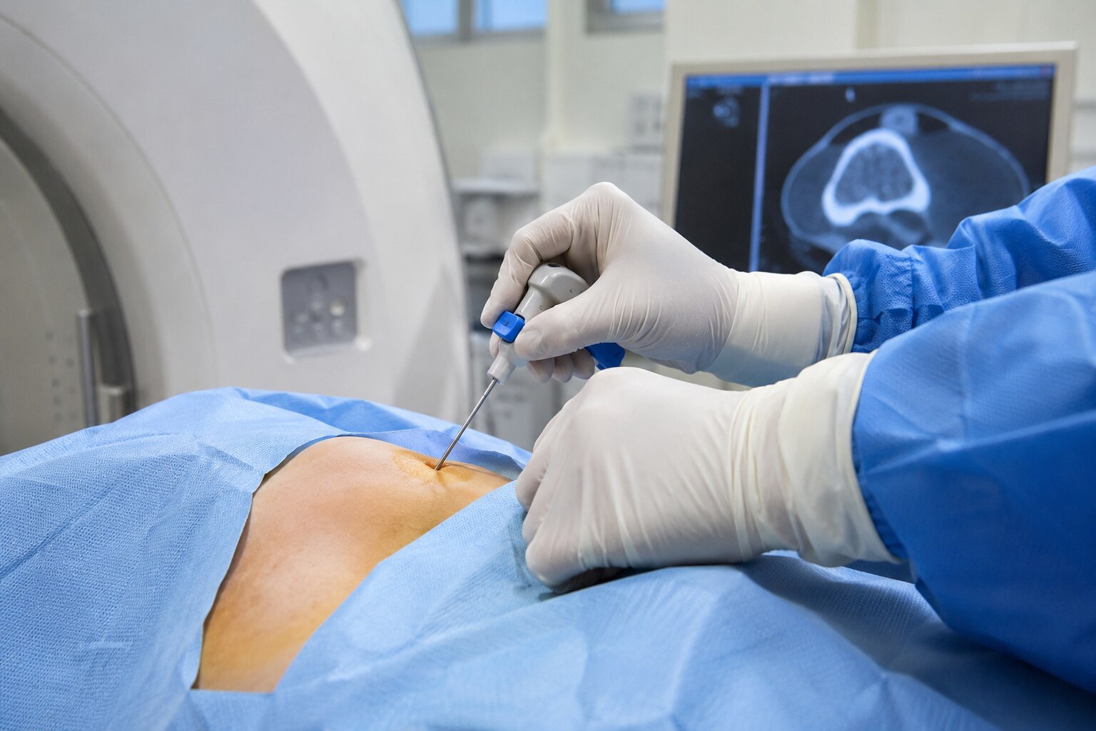
A needle biopsy describes how tissue is removed: a needle passes through skin into the lesion. Image guidance describes how the doctor positions that needle, using CT, ultrasound, fluoroscopy or another imaging method. Therefore, image-guided biopsy does not replace needle biopsy; it is a way to perform one more accurately.
If you are comparing needle biopsy or image guided biopsy for bone tumors in Mumbai, ask whether the planned procedure is image-guided core needle biopsy. “Core” describes the solid tissue cylinder collected, while “image-guided” describes needle placement.
| Option | What it means | Main value |
|---|---|---|
| Needle biopsy | Tissue is sampled through a needle | Less invasive sampling than open surgery |
| Image-guided biopsy | Imaging directs the needle into the target | Avoids nearby structures and improves targeting |
| Core needle biopsy | A cutting needle removes tissue cylinders | Provides architecture for tumor classification |
A correctly placed needle can still produce an inadequate result. The sample might contain dead tissue, blood, reactive bone, or too little viable tumor. A nondiagnostic report does not prove that cancer is absent; the next step may be repeat sampling or open biopsy.
For bone tumor diagnosis in Mumbai, the biopsy route should be planned after reviewing the radiographs and MRI. The orthopedic-oncology team performing definitive surgery should choose the entry point because the biopsy track may need removal with the tumor.
A musculoskeletal pathologist should interpret the specimen alongside imaging and clinical history, with cultures, stains or molecular tests added when the diagnosis requires them.
How doctors choose CT, MRI, ultrasound or fluoroscopy for a bone lesion
The safest path is the one that reaches viable tumour while avoiding major nerves, blood vessels, joints and organs. Doctors choose the modality after reviewing radiographs and MRI, then plan the needle track so the definitive tumour surgeon can remove it later.
| Imaging method | What it shows best | When it is chosen |
|---|---|---|
| CT | Cortical bone, mineralisation, deep anatomy and needle position | Small, deep, pelvic, spinal or anatomically complex lesions |
| MRI | Marrow involvement, soft-tissue extension and relationship to nerves or vessels | Mapping the lesion and, in selected centres, guiding the biopsy |
| Ultrasound | Superficial soft tissue and vessels in real time | Accessible lesions or a soft-tissue component visible on ultrasound |
| Fluoroscopy | Live X-ray views of bones and instruments | Lesions with clear bony landmarks, when limited real-time guidance is sufficient |
CT is particularly useful when the lesion is deep or hidden behind bowel, muscle or other bone. Ultrasound avoids radiation, but it cannot see through intact cortical bone and is unsuitable when the target is deep without a visible soft-tissue component. Fluoroscopy is quick, but provides less cross-sectional detail than CT.
MRI guidance is reserved for selected targets because equipment access and needle compatibility can limit its use.
Ask who will plan the route, whether MRI findings and pathology will be reviewed together, and what happens if the sample misses viable tumour. A correctly positioned needle can still collect necrotic tissue, blood or reactive bone.
For anyone comparing image guided bone biopsy in Mumbai, the team’s experience with the lesion’s location and planned surgery matters as much as the scanner.
Core needle, fine-needle, incisional or open biopsy: which sample is appropriate?
Core needle biopsy is preferred when the pathologist needs tissue architecture, not only loose cells. A spring-loaded needle removes cylinders from viable tumor, allowing assessment of subtype and grade plus immunohistochemical or molecular tests.
Fine-needle aspiration removes cells into a syringe; it can help with selected lesions but may not distinguish a primary bone tumor from metastasis, infection or reactive tissue.
| Option | Sample obtained | Main strength | Main limitation |
|---|---|---|---|
| Core needle biopsy | Cylinders of tissue | Architecture, grading, immunohistochemistry and molecular testing | Can miss tumor if it samples necrosis, blood or reactive bone |
| Fine-needle aspiration | Individual cells and fluid | Quick cytology for selected lesions or suspected recurrence | Often too little structure to classify a primary bone tumor |
| Incisional biopsy | A piece through a surgical incision | Larger tissue sample when needle cores are inadequate | More pain, wound risk and a surgical scar |
| Open or excisional biopsy | Surgical tissue removal; sometimes the whole lesion | Useful when needle sampling remains nondiagnostic or the lesion is small and removable | May complicate later tumor surgery; excision is not automatically curative |
A correctly placed needle can still produce a nondiagnostic result. Ask whether the target contains viable tumor, what happens if the cores are inadequate, and whether imaging and pathology will be reviewed together.
For a suspected primary bone tumor, the surgeon who may perform definitive treatment should plan the biopsy track because that tract may need removal with the tumor.
This coordination matters when arranging a bone cancer biopsy in Mumbai or comparing centres offering bone tumor diagnosis in Mumbai. A core biopsy diagnoses; it does not remove or cure the tumor.
Why the biopsy route must be planned with definitive tumor surgery
The entry point is chosen for the operation that may follow, not simply for the shortest route to the lesion. After radiographs and MRI are reviewed, the team performing definitive tumor surgery plans a path that can be removed with the tumor.
A poorly placed tract can contaminate uninvolved tissue compartments and force a wider resection.
The plan accounts for:
- Suspected diagnosis: a core must target viable tumor rather than necrotic tissue and provide enough architecture for grading, immunohistochemistry or molecular testing.
- Lesion location: a deep pelvic lesion, cortical lesion or tumor with soft-tissue extension may require CT guidance and a carefully selected approach through one safe compartment.
- Future operation: the tract is generally longitudinal, kept within the planned surgical field and kept away from a joint when possible. The bone involved and the planned reconstruction can change that route.
The number of samples depends on the pathology questions and the amount of tissue safely obtainable. One small fragment may be inadequate for diagnosis, grading and additional tests; several cores from the solid, enhancing portion often give the laboratory a better chance of answering all three.
The biopsy team should agree in advance on specimen handling if infection, lymphoma or another unusual diagnosis is suspected.
If you are comparing needle biopsy for bone tumor in Mumbai or researching bone tumor diagnosis in Mumbai, ask who has approved the tract, how many cores are planned, and what happens if the sample is inadequate. The needle samples tissue; it does not remove or cure the tumor.
What happens before, during and after the biopsy—and what should you ask in Mumbai?
Before the appointment, confirm that the orthopedic-oncology team has reviewed the radiographs and MRI, chosen the entry route, and planned for the needle track to be removed during definitive surgery. Ask whether the sample will be core tissue, how many cores are needed, and what happens if the first sample is inadequate.
- Before: disclose blood thinners, diabetes, allergies, pregnancy possibility and previous bleeding problems. Confirm fasting instructions, local anaesthetic versus sedation, medication changes, transport home and expected observation time. Ask who will perform the procedure and who will coordinate pathology.
- During: imaging guides placement, but accurate placement does not guarantee diagnosis. Necrotic tissue, haemorrhage, reactive bone or too little tissue can produce a nondiagnostic result. Ask whether tissue is being allocated for histology, immunohistochemistry, molecular testing or microbiology if infection is a possibility.
- After: expect soreness or bruising; seek urgent care for worsening pain, fever, persistent bleeding, swelling or new weakness. Ask when the pathology report is expected, whether the radiologist, pathologist and surgeon will review the images together, and whether a nondiagnostic result means repeat needle biopsy or open biopsy—not that cancer is absent.
For image guided bone biopsy in Mumbai or a bone cancer biopsy in Mumbai, ask which pathology laboratory handles the specimen and whether the surgeon plans the tract. Dr Mishil Parikh is an orthopedic oncologist, so ask him specifically how the proposed route fits the eventual operation.
Discuss staging separately: biopsy confirms tissue type; MRI, chest imaging and other tests assess spread. This is the key distinction when comparing needle biopsy or image guided biopsy for bone tumors in Mumbai.
Related services
Image Guided Biopsy Image-guided biopsy is a procedure that uses medical imaging techniques, such as ultrasound, computed tomography (CT), magnetic resonance imaging... View service → |
Core Needle Biopsy Core needle biopsy is a minimally invasive diagnostic procedure used to obtain tissue samples from suspicious areas in the body for further... View service → |
Frequently asked questions
What is the difference between a needle biopsy and an image-guided biopsy?
A needle biopsy describes how tissue is removed through the skin. An image-guided biopsy uses CT, MRI, ultrasound or fluoroscopy to position that needle accurately.
How do doctors choose CT, MRI, ultrasound or fluoroscopy for a bone lesion?
CT is useful for deep lesions and complex bony anatomy, ultrasound for accessible soft-tissue targets, MRI for soft-tissue relationships, and fluoroscopy for real-time guidance during selected procedures.
Which biopsy type is appropriate for a bone tumor?
A core needle biopsy often provides tissue architecture, while fine-needle, incisional or open biopsy is chosen when the diagnosis requires a different or larger sample.
Why must the biopsy route be planned with definitive tumor surgery?
The route must allow the biopsy tract to be removed during definitive surgery. An incorrectly placed tract can contaminate additional tissue compartments and complicate treatment.
What should you ask before a bone tumor biopsy in Mumbai?
Ask which imaging method and biopsy type are planned, who will review the pathology, how the route relates to surgery, and what preparation and follow-up you need.
Keywords