A needle biopsy involves more than placing a needle into a lump: the scan sequence, needle route, tissue quantity, medicines, and laboratory tests all affect the result. By the end, you will know how to prepare, what the appointment feels like, how long answers can take, and what to do if the pathology result does not fit the scan.
Key takeaways
- Core biopsy preserves tissue architecture for subtype, grade, and additional testing.
- Tell your team about blood thinners, allergies, and previous biopsy results.
- The needle route must be planned so it can be removed during definitive surgery.
- Ask whether the pathology result matches the scan, examination, and biopsy target.
Why a core biopsy is chosen over other biopsy types
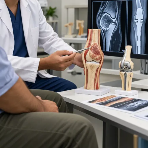
A core needle biopsy is chosen because it removes small cylinders of tissue, not just loose cells. Those cores preserve tissue architecture, allowing a specialist to assess whether a mass is a sarcoma, identify its subtype, estimate its grade, and obtain material for immunohistochemistry or molecular testing.
Image guidance helps target viable areas, and several cores may be taken when a mass contains necrosis, bleeding, or calcification.
| Biopsy type | What it provides | Main limitation or trade-off |
|---|---|---|
| Core needle biopsy | Tissue cylinders with cells and architecture | Requires carefully planned needle placement |
| Fine-needle aspiration | Cells drawn through a thin needle | Often lacks architecture and enough tissue for sarcoma grading or molecular tests |
| Incisional biopsy | A surgically removed portion of the mass | Larger wound and greater risk of disrupting tissue planes |
| Excisional biopsy | The entire mass | May be inappropriate for a large or deep tumor because removal can compromise definitive surgery |
Fine-needle aspiration can help evaluate some lumps, but its cells alone may not distinguish closely related tumor types. A core needle biopsy usually gives the pathology team more evidence without the wound and recovery of an open operation.
An incisional biopsy is reserved for situations where needle samples are inadequate or unsafe. An excisional biopsy is generally suitable only for a small, superficial lesion that can be removed with planned margins.
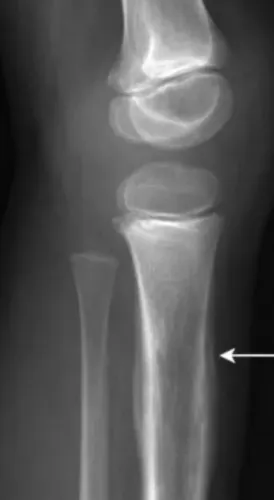
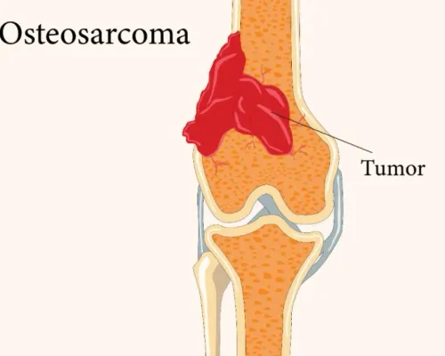
For a suspected bone or soft-tissue sarcoma, the core tract must be placed in a straight path that the orthopedic tumor surgeon can remove during later surgery.
How doctors choose the safest biopsy route
A suspicious soft-tissue mass usually needs MRI before biopsy. MRI maps the tumor’s compartments and shows whether it touches a nerve, blood vessel, joint, or bone; entering before that map exists can cross tissue planes that later need to remain clear.
| Situation | Imaging used to choose entry | How the route is planned | Future-surgery concern |
|---|---|---|---|
| Superficial, clearly visible mass | Ultrasound, often with MRI | Enter through the shortest safe path | Keep the biopsy track within tissue that can be removed |
| Deep or anatomically complex mass | MRI for mapping; CT-guided biopsy for needle placement | Avoid vessels, nerves, joints, and uninvolved muscle compartments | Prevent contamination of a second surgical compartment |
| Bone lesion or pelvic tumor | CT-guided biopsy, with MRI or other scans for extent | Select a straight approach through the least disruptive bone and soft tissue | Place the track where the tumor surgeon can remove it with the specimen |
| Mass near major vessels or nerves | MRI, CT, or both | Choose an angle that avoids the neurovascular bundle | Do not leave a track that would require sacrificing a critical structure |
The biopsy track is not an incidental puncture. Surgeons usually mark its path so the entire channel, including the skin opening, can be removed during definitive surgery. Tumor-cell implantation along the track is uncommon with planned image-guided technique, but a poorly placed track can complicate an otherwise manageable operation.
In sarcoma biopsy planning, coordinate the route with the orthopedic tumor surgeon who may perform the definitive procedure. Dr Mishil Parikh uses that surgical question to help determine where the needle should enter, not merely where the mass is easiest to reach.
Related services
Core Needle Biopsy Core needle biopsy is a minimally invasive diagnostic procedure used to obtain tissue samples from suspicious areas in the body for further... View service → |
Image Guided Biopsy Image-guided biopsy is a procedure that uses medical imaging techniques, such as ultrasound, computed tomography (CT), magnetic resonance imaging... View service → |
What to do before and what happens during the appointment
Your biopsy preparation begins with a medication and health review. Tell the team about blood thinners, antiplatelet drugs, bleeding disorders, allergies, diabetes, and possible pregnancy. Do not stop an anticoagulant on your own; the team will decide whether the dose needs adjustment. Bring relevant scans, reports, and medication details.
- Follow the appointment instructions. If sedation is planned, you may need to fast for a specified period and arrange an adult to take you home. Ask when you can resume food, drink, and regular medicines.
- Change into suitable clothing and remove anything that obstructs the biopsy area. You will give consent after the doctor explains the route, benefits, risks, and alternatives.
- Lie in the position chosen from your scans. The team cleans your skin and covers it with sterile drapes.
- You receive a local anaesthetic through a small injection. It can sting briefly, then numb the skin and deeper tissues.
- During each needle pass, expect pressure, pushing, or a short-lived ache rather than a sharp cut. Bone lesions can hurt more when the needle crosses the hard outer cortex; tell the team if discomfort becomes significant.
- After the samples are taken, the needle site receives a dressing. Staff observe you before discharge.
Mild soreness or a small bruise is common immediately afterward. Contact the medical team promptly for increasing swelling, ongoing bleeding, fever, worsening pain, new numbness or weakness, or a cold or discoloured limb.
What happens to the tissue after the needle comes out
Each core is placed in a formalin container that preserves its structure during transport. If molecular testing is planned, the team may reserve part of the specimen in another medium or retain unstained sections, because processing all tissue at once can leave too little material for later tests.
In the laboratory, pathology processing usually follows these steps:
- The laboratory records the specimen, measures it, and selects representative pieces.
- The tissue is processed into paraffin blocks and cut into very thin sections.
- The pathologist examines the sections under a microscope with routine stains.
- Additional sections may undergo immunohistochemistry, which uses antibody-based stains to identify proteins that help classify the tumor.
- Cytogenetic tests or molecular testing may examine chromosome changes, gene fusions, mutations, or other features.
A preliminary microscopic impression can therefore arrive before the final diagnosis. The pathologist must interpret the tissue alongside your scans and clinical history, and a small or necrotic sample may not contain enough viable tumor for every test. Bone samples can also require decalcification, a treatment that adds time and can affect some molecular assays.
If results do not match the imaging, the case may need specialist review, extra stains, or testing on additional tissue. Ask when the final integrated report is expected rather than relying on the first verbal update.
How to read the result and recognise a mismatch
A pathology report may identify a benign lesion, a malignant tumor, infection, or a specific tumor type and grade. It is read alongside your scans, examination findings, and medical history; a result cannot be judged from the microscope description alone.
A “non-diagnostic biopsy” means the tissue does not answer the clinical question. The sample may contain blood, necrotic tissue, calcification, scar, or too little viable tumor. It does not prove that the mass is benign.
Ask what the result means and what happens next:
- If the tissue is diagnostic, your team uses the tumor type, grade, and imaging to plan treatment. Additional immunohistochemistry, cytogenetic testing, or next-generation sequencing can extend the final report.
- If the result is non-diagnostic, the specialist reviews the needle path, sample quality, and scans. The team may recommend a repeat image-guided biopsy or an open biopsy.
- If pathology suggests a benign lesion but imaging shows aggressive bone destruction, rapid growth, or invasion of nearby structures, treat that disagreement as unresolved. The same applies when a report says infection but the clinical and imaging picture suggests a malignant tumor.
- A sarcoma-focused multidisciplinary team compares the slides, scans, and clinical history. It may request another pathology opinion or obtain more tissue before treatment.
Do not proceed to definitive tumor surgery based on a result that does not fit the imaging. A second biopsy can prevent both undertreatment and an unnecessarily extensive operation.
Frequently asked questions
Why is a core biopsy used instead of a fine-needle aspiration?
A core biopsy removes tissue cylinders that preserve architecture. That helps specialists assess sarcoma subtype and grade and perform immunohistochemistry or molecular testing.
How do doctors choose the safest biopsy route?
They use imaging, the mass location, nearby nerves and blood vessels, and the planned definitive surgery. The route should avoid contaminating tissue that would not be removed later.
What should I do before a needle biopsy?
Give the team a complete list of medicines, especially blood thinners, plus allergies, bleeding problems, and earlier imaging or biopsy reports. Follow the stated instructions about eating, drinking, and arranging transport.
What happens to the tissue after the needle comes out?
The laboratory fixes, processes, and examines the cores. A specialist may add immunohistochemistry or molecular tests when the initial appearance does not identify the tumor precisely.
What does a mismatch between the biopsy result and the scan mean?
A mismatch means the pathology does not adequately explain the imaging, examination, or clinical behaviour. Ask whether the sample came from the correct target and whether repeat biopsy or specialist review is needed.