A mole that changes, bleeds, itches, or looks unlike your other marks deserves a closer look, but no checklist can diagnose melanoma by appearance alone. You will learn which changes warrant routine or prompt assessment, what to record before your appointment, and how clinicians move from examination to dermoscopy, biopsy, pathology, and staging.
Key takeaways
- Arrange assessment for a changing, asymmetric, multicoloured, bleeding, or unusual spot.
- Check palms, soles, nails, scalp, eyes, and other less visible sites.
- Bring dated photographs and note changes, symptoms, sun exposure, and family history.
- Only examination and biopsy can confirm melanoma; appearance alone cannot diagnose it.
Use ABCDE, the ugly-duckling sign, and EFG to check a changing spot
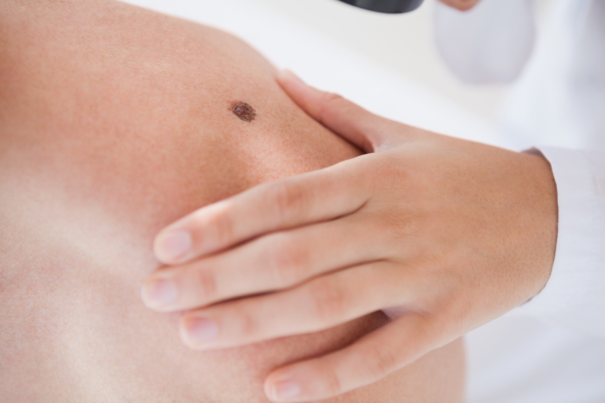
Use the ABCDE rule to spot melanoma warning signs, but treat it as a reason to arrange assessment, not as a diagnosis. Check a mole in good light and compare it with earlier photographs.
1. Look for asymmetry: one half does not resemble the other. An irregular border melanoma may have an uneven, scalloped, notched, or poorly defined edge. Uneven colour, including several shades of brown, black, red, blue, or white, is another warning sign.
2. Check diameter. A suspicious spot is usually greater than 6 mm, about the size of a pencil eraser, but melanomas can be smaller than 6 mm. Evolution matters more than measurement: report any change in size, shape, colour, symptoms, or surface.
3. Find the ugly duckling. A mole that looks noticeably different from your other moles can be suspicious even when it lacks several ABCDE features. Also arrange assessment for a new spot after age 30, a sore that does not heal, spontaneous bleeding, crusting, persistent itching or tenderness, or a rapidly enlarging lump.
For a rapidly growing nodular pattern, use EFG: elevated, firm, and growing. Nodular melanoma can appear relatively symmetric and evenly coloured, so ABCDE alone can miss it.
These findings also occur in harmless conditions, infections, or injuries. They trigger professional assessment; they do not prove melanoma. A clinician may need dermoscopy or a biopsy to identify the cause.
Check less obvious sites and avoid diagnosing benign look-alikes by sight
Melanoma in dark skin can arise on the palms, soles, under fingernails, the scalp, inside the mouth, or in the eye—not only on sun-exposed skin. Acral melanoma may appear as a new or changing irregular brown or black patch.
Nail melanoma may form a longitudinal dark band; pigment spreading onto the surrounding skin is called Hutchinson sign.
A harmless-looking lesion can still need examination because infection, trauma, and benign growths change, while melanoma can look ordinary.
| Finding | Why it may seem harmless | Why sight is not enough |
|---|---|---|
| Seborrhoeic keratosis | Waxy, stuck-on, or dark surface | Melanoma can mimic a pigmented growth |
| Dermatofibroma | Firm, small, often longstanding bump | A changing firm lesion needs assessment |
| Ordinary mole | Even colour and familiar shape | Melanoma can resemble a routine mole |
| Infected spot | Redness, tenderness, crust, or drainage | Infection can obscure an underlying tumour |
| Blood blister | Dark, raised mark after friction or injury | Trauma does not prove the pigment is blood |
Do not dismiss a lesion because it is raised, dark, tender, or located where sun exposure is limited. Arrange professional assessment for a new or changing patch, a dark nail streak, or a lesion that bleeds, crusts, itches, hurts, or fails to heal.
Visual comparison can raise concern, but dermoscopy and, when indicated, biopsy determine what it is.
Choose the right appointment and prepare a useful history
Book a routine dermatology appointment for a new or changing skin lesion, an ugly duckling, a new dark nail streak, or persistent itch or tenderness. Use this guide to choose how quickly to act:
| Situation | Action | Why |
|---|---|---|
| New or changing spot, ugly duckling, dark nail streak, persistent itch or tenderness | Arrange a dermatology visit | These changes need professional assessment |
| Spontaneous bleeding, a bleeding mole, rapid enlargement, significant pain, ulceration, or a sore that does not heal | Seek prompt assessment | Waiting can delay diagnosis |
Prepare a useful history before you go:
- Record when the lesion appeared.
- Write the sequence of changes in size, shape, colour, or surface.
- Note bleeding, crusting, itch, pain, or tenderness.
- List personal and family history of melanoma, previous skin cancers, immune problems, sun exposure, and tanning-bed use.
- Take dated skin lesion photographs with a ruler beside the lesion for scale.
Photographs help show evolution, especially when appointments are weeks apart, but they do not replace examination. A clinician must assess the lesion directly, often with dermoscopy, because infection, trauma, and benign growths can mimic melanoma.
If you cannot obtain a prompt dermatology appointment and the lesion is bleeding without injury, rapidly enlarging, ulcerated, or significantly painful, contact a doctor or urgent-care service.
What happens during examination, dermoscopy, and biopsy
A suspicious skin change needs tissue diagnosis when its history, examination, or dermoscopic pattern remains concerning; appearance alone cannot decide. The clinician asks when it appeared, how it evolved, and whether it bleeds, crusts, itches, hurts, or feels tender, while reviewing previous skin cancers, sun exposure, family history, and immune status.
They inspect the entire skin surface, compare lesions, and palpate regional lymph-node basins. The scalp, nails, soles, and mucosal surfaces are examined when the history or lesion pattern calls for it. Dermoscopy reveals structures and colours not visible to the unaided eye, but it does not replace histopathology.
The definitive test is histopathology from a melanoma biopsy. When feasible, clinicians generally prefer complete excisional biopsy of the clinically suspicious lesion with a narrow margin. A superficial shave that transects the lesion can leave the deepest area unsampled and prevent accurate depth measurement.
Key pathology-report details include:
- Breslow thickness, measured in millimetres
- Ulceration
- Mitotic activity, where reported
- Microsatellites
- Whether the margins are involved
How confirmed melanoma is staged—and when other specialists become involved
1. Pathology starts melanoma staging, but the first biopsy is not the final cancer operation. A complete excisional biopsy with a narrow margin confirms the diagnosis and measures Breslow thickness.
If melanoma is confirmed, a later wide local excision removes additional normal-looking skin; its margin is selected according to tumour thickness and anatomical site, not by simply repeating the biopsy margin.
2. A sentinel lymph-node biopsy is a staging procedure, not a test for every suspicious mole. The team considers it from the confirmed thickness and adverse features, especially ulceration; very thin melanomas without high-risk findings often do not require it. The result helps determine prognosis and further treatment.
3. CT, PET, MRI, and bone scans are chosen from the stage, symptoms, examination findings, or concern for spread. They are not automatic tests for every new melanoma, particularly when the disease appears localized on skin assessment and pathology.
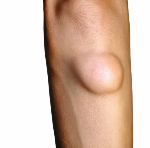
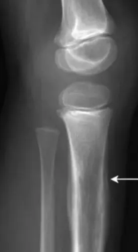
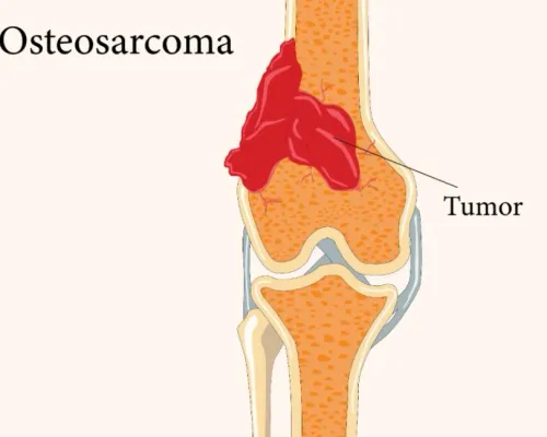
4. Persistent focal bone pain, swelling, weakness, or a pathologic fracture in someone with known or advanced melanoma needs coordinated oncology and orthopedic tumour assessment. These findings can prompt investigation for melanoma bone metastasis, with imaging and biopsy planned by the treating specialists.
In Mumbai, Dr Mishil Parikh’s musculoskeletal-oncology practice can assess suspected tumour involvement of bone or soft tissue alongside the oncology team; it is not a substitute for diagnosing the original skin lesion.
Related service
Skin Cancers Skin cancer refers to the abnormal growth of skin cells, typically as a result of damage to the DNA of the skin cells caused by exposure to... View service → |
Frequently asked questions
What melanoma warning signs should you check at home?
Use ABCDE, the ugly-duckling sign, and EFG: look for asymmetry, irregular borders, uneven colour, growth, change, firmness, and elevation.
Where can melanoma appear besides commonly exposed skin?
Check the palms, soles, spaces between toes, fingernails, toenails, scalp, eyes, mouth, and genital skin.
What happens during a melanoma assessment?
A doctor examines your skin, may use dermoscopy to magnify structures, and removes a suspicious area for biopsy when needed.
How is confirmed melanoma staged?
Staging uses the biopsy findings and, when indicated, imaging, lymph-node assessment, or other tests; specialists then plan treatment together.