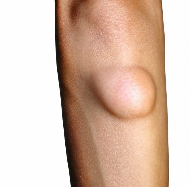
A growing lump is not automatically sarcoma, but certain features make waiting for it to disappear a poor strategy. You will learn which changes need prompt assessment, how common explanations can mislead, what an examination and imaging work-up involve, and why biopsy planning matters before removal.
Key takeaways
- Arrange prompt assessment for an enlarging, fixed, deep, painful, or 5-centimetre lump.
- A lump’s appearance cannot reliably distinguish sarcoma from lipoma, strain, or bruise.
- Specialists assess size, depth, mobility, tenderness, growth, and nearby nerves or blood vessels.
- Ask why imaging and a planned biopsy should come before surgical removal.
Which lump changes warrant prompt specialist assessment?
An enlarging lump, a mass about 5 centimetres or larger, a deep mass beneath the fascia, a fixed lump, or pain warrants prompt medical assessment. A 5-centimetre soft tissue mass is roughly the size of a golf ball. These features raise concern for soft tissue sarcoma, but none diagnoses cancer on its own.
| Feature | What it means | What to do |
|---|---|---|
| Enlarging lump | Growth matters even when it is slow and painless | Arrange prompt assessment |
| 5 cm or larger | About the size of a golf ball | Request medical review |
| Deep or fixed lump | It may lie beneath fascia or attach to nearby tissue | Seek specialist assessment |
| Painful lump | Pain can reflect pressure or involvement of nearby structures | Arrange prompt review |
| Superficial, mobile lump | Mobility does not prove it is benign; some sarcomas begin in subcutaneous tissue | Do not rely on touch alone |
Rapid growth deserves faster attention, but slow growth does not make a mass safe. Some sarcomas enlarge gradually and remain painless, so do not wait for pain, bruising, or skin changes. A painless lump that keeps growing or feels deep still needs review.
Seek urgent care for severe or rapidly worsening pain, fever, skin breakdown, sudden marked swelling, persistent numbness, weakness, loss of limb function, or signs of impaired circulation. These symptoms can indicate infection, bleeding, or compression of a nerve or blood vessel and require immediate assessment even when cancer is not the cause.
How can you tell a concerning mass from a lipoma, strain, or bruise?
You cannot reliably tell a sarcoma from appearance or touch alone. A soft, mobile lump fits a common lipoma, but mobility does not rule out malignancy; lipoma versus sarcoma requires clinical assessment and sometimes imaging or biopsy. A muscle strain usually improves with time and activity modification.
A post-injury hematoma or bruise should gradually shrink, not continue enlarging.
Trauma can draw attention to a pre-existing mass rather than cause it. Re-examine any presumed bruise, hematoma, or post-traumatic swelling that persists, grows, or fails to follow the expected recovery pattern. An abscess or cyst may be tender, warm, red, or fluctuant, but persistent or recurring findings need assessment.
Before review, record:
- The date you first noticed the lump, its location, length and width, whether it is changing, and how quickly it has grown
- Any injury, injection, prior cancer, or radiation exposure
- Fever, weight change, night symptoms, numbness, weakness, or reduced movement
Take dated photographs only to record growth. Do not squeeze, drain, massage, or repeatedly press the mass, since this can worsen irritation and make changes harder to judge. Arrange review when the history does not fit a straightforward resolving injury or infection, even if the lump is painless.
What does a specialist examination look for?
A specialist examination locates the mass precisely and compares it with the opposite side. The clinician measures it, checks whether it moves beneath the skin or with muscle contraction, and determines whether it is a superficial versus deep mass in relation to the fascia.
The examination checks:
- Firmness, tenderness, fixation, skin colour, warmth, ulceration, visible veins, and changes after movement
- Whether a superficial lump can be inspected and palpated directly
- Whether a deep mass could involve muscle, fascia, a nerve, a blood vessel, or bone, making its edges difficult to define by touch
- Neurological findings, including sensation, muscle strength, reflexes, and limb function when nerve compression is possible
- Circulation and lymph nodes, including pulses, limb temperature, swelling, other circulation changes, and regional lymph-node enlargement
Tell the clinician how quickly the mass has grown and mention every treatment already attempted, including antibiotics, drainage, massage, or surgery. A normal-looking skin surface does not exclude a deep tumour, and a normal plain X-ray does not exclude a soft-tissue sarcoma.
The examination determines whether you need observation, ultrasound, urgent referral, or a specialist-led imaging and biopsy plan. Seek prompt assessment for new numbness, weakness, reduced limb function, marked swelling, or circulation changes, because these findings can indicate compression of a nearby nerve or vessel.
Which tests come next, and why should biopsy precede removal?
Ultrasound is often the first test for a new superficial mass. It can show whether the lesion is solid or cystic, its depth, blood flow, and relationship to nearby structures. A reassuring ultrasound does not override continued growth, deep location, fixation, or other concerning findings.
| Test | What it shows | What it cannot establish |
|---|---|---|
| Ultrasound | Solid versus cystic structure, depth, blood flow, and nearby anatomy | A reassuring result cannot rule out sarcoma when warning features persist |
| MRI with contrast | Local extent against fascia, muscle, nerves, vessels, and bone | It maps disease but cannot replace tissue diagnosis |
| Plain X-ray | Calcification, bone involvement, or another explanation for symptoms | A normal image cannot rule out soft-tissue sarcoma |
MRI with contrast is the preferred local study for most suspected soft-tissue sarcomas. If sarcoma is possible, refer to a specialist team before definitive excision. The team commonly plans a core-needle biopsy after imaging, with the biopsy route positioned so the tract can be removed with the tumour during surgery.
Properly performed needle biopsy does not generally spread sarcoma. Poor planning creates the greater practical risk. Removing a lump without this plan, sometimes called a whoops excision, can contaminate surrounding tissue and require wider re-excision or additional treatment.
When sarcoma is diagnosed or strongly suspected, chest imaging is included because the lungs are a common site of spread.
How should you arrange the referral and prepare for the visit?
Contact a doctor promptly when a lump is enlarging, about 5 centimetres or larger, deep to the fascia, fixed, or painful. Request sarcoma specialist referral to a musculoskeletal tumour service before anyone attempts definitive removal.
1. Write the history before appointment. Record the date you first noticed the lump, its measurements, growth rate, injury or injection history, previous cancer, radiation exposure, and any pain, fever, skin change, numbness, weakness, swelling, or loss of function.
2. Bring imaging reports, scan images if available, and a complete medicine list. Mark blood thinners clearly because they affect biopsy planning and bleeding risk.
3. Ask where the mass sits in relation to the fascia and whether ultrasound or MRI is the appropriate next test. Ultrasound often assesses a superficial lump; MRI maps a suspected tumour against muscle, nerves, vessels, and bone.
4. Ask who will plan a biopsy if one is needed, which biopsy type is proposed, and where the needle will enter before agreeing to excision. An unplanned removal can leave tumour cells in surrounding tissue and complicate further surgery.
Imaging, pathology, staging, radiotherapy decisions, and surgery are interdependent, so specialist coordination matters. An orthopaedic tumour surgeon such as Dr Mishil Parikh can assess bone and soft-tissue tumour concerns and help decide between surveillance, biopsy, and tumour-focused treatment.
Choose urgent care instead of waiting for a routine appointment if severe pain, fever, skin breakdown, new weakness or numbness, rapidly increasing swelling, or loss of limb function develops.
Related services
Soft Tissue Tumor A soft tissue tumor refers to an abnormal growth of cells that occurs in the soft tissues of the body. View service → |
Image Guided Biopsy Image-guided biopsy is a procedure that uses medical imaging techniques, such as ultrasound, computed tomography (CT), magnetic resonance imaging... View service → |
Frequently asked questions
Which soft-tissue lump changes warrant prompt specialist assessment?
Seek prompt assessment for an enlarging lump, a mass about 5 centimetres or larger, a deep mass beneath the fascia, a fixed lump, or pain. These signs raise concern but do not diagnose sarcoma.
How can you tell a concerning mass from a lipoma, strain, or bruise?
You cannot reliably distinguish them by touch or appearance alone. A clinician considers the lump’s growth, size, depth, fixation, pain, and examination findings, then uses imaging when needed.
What does a specialist examination look for?
The specialist checks the mass’s size, depth, mobility, tenderness, rate of growth, relationship to nearby muscles, and possible involvement of nerves or blood vessels.
Which tests come next, and why should biopsy precede removal?
Imaging helps define the mass and guide planning. A planned biopsy should precede removal when sarcoma is suspected so the biopsy path and surgery can be coordinated safely.
How should you arrange the referral and prepare for the visit?
Bring referral letters, scan reports and images, pathology results, a medication list, and a timeline recording when the lump appeared, changed, or became painful.