The right sample depends on whether doctors are investigating systemic myeloma, a solitary bone lesion, or a fracture weakened by an unknown tumour. By comparing marrow biopsy, lesion biopsy, needle techniques and open surgery, you can understand which test fits the scans, the urgency and the treatment plan.
Key takeaways
- Myeloma usually requires bone-marrow aspirate and trephine biopsy.
- Use imaging to select the safest target and needle route.
- Treat a suspected pathological fracture as an urgent orthopaedic problem.
- Ask how the sample will be labelled, transported and tested.
Which tissue sample is needed when myeloma is suspected?
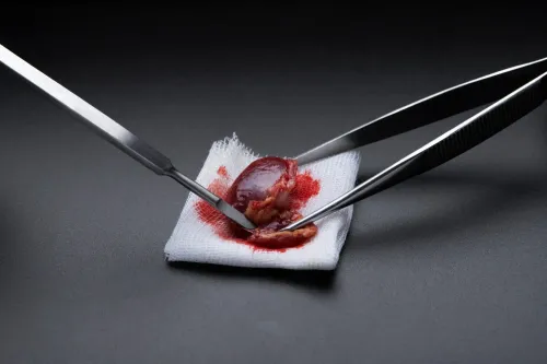
Multiple myeloma is usually confirmed with a bone-marrow aspirate and trephine biopsy, not automatically by sampling the painful bone lesion. The marrow sample measures plasma cells and supports flow cytometry, plasma-cell immunophenotyping, immunohistochemistry and, when indicated, cytogenetic or FISH studies.
| Option | What it samples | When it applies |
|---|---|---|
| Marrow aspirate and trephine | Liquid marrow and a solid marrow core | Suspected systemic myeloma |
| Targeted core biopsy | The abnormal bone or soft-tissue mass | Atypical mass, uncertain diagnosis or suspected solitary plasmacytoma |
| FNA | Loose cells only | Supplement to core tissue, not usually sufficient alone |
| Open or incisional biopsy | A larger surgical tissue sample | Repeated nondiagnostic needle samples or inaccessible lesions |
| No immediate biopsy | No new tissue initially | Blood, urine, marrow and imaging already establish myeloma |
Diagnosis also uses serum protein electrophoresis, immunofixation, serum free light chains, urine studies, whole-body imaging and plasma-cell testing. A lesion biopsy can distinguish myeloma from metastatic carcinoma, lymphoma, sarcoma or infection, while a pathological fracture can also result from benign bone disease.
Core tissue preserves architecture for histology and immunohistochemistry, so it is usually more useful than FNA alone. For a core needle biopsy for myeloma in Mumbai or a biopsy for bone lesion in Mumbai, state “suspected myeloma” on the request. This lets pathology reserve tissue for flow cytometry and molecular studies.
A separate lesion biopsy may be unnecessary when systemic findings are conclusive.
How imaging determines the biopsy target and needle route
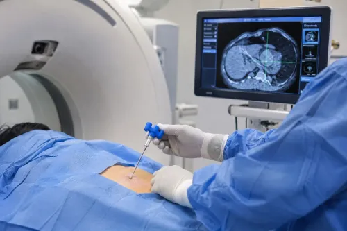
Clinicians do not simply sample the most visible part of a lesion. They compare radiographs, CT, MRI and PET-CT to identify viable tumour, then target an accessible soft-tissue component when it represents the lesion. They avoid necrotic, infected, haemorrhagic or previously contaminated tissue because it can produce a misleading or nondiagnostic result.
| Guidance | Best target | What it shows |
|---|---|---|
| CT-guided biopsy | Deep, pelvic, spinal, sclerotic or anatomically complex lesions | The intraosseous target and the planned path around vessels, nerves and organs |
| Ultrasound-guided biopsy | Superficial lesions or an accessible soft-tissue extension | The needle in real time and nearby superficial vessels; it may not show the bone target adequately |
The radiologist, orthopaedic oncologist and pathologist should agree on the target and route before the procedure. The needle track must remain within the planned definitive surgical field so the surgeon can remove it with the tumour. An incorrectly placed track can contaminate extra tissue planes and compromise limb-sparing surgery.
A small lesion, heavily sclerotic bone or mostly necrotic mass carries a higher sampling-error risk. If the pathology conflicts with the scans or clinical findings, repeat image-guided core biopsy, a different target or open biopsy may be necessary.
There is no single best needle biopsy option in Mumbai; the right choice depends on imaging, anatomy and laboratory needs. Anyone arranging a biopsy for bone lesion in Mumbai should ask who planned the track and what happens after a nondiagnostic sample.
What changes when a pathological fracture needs urgent care?
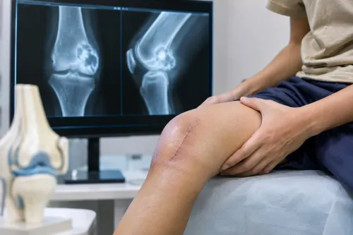
A biopsy diagnoses the cause of a pathological fracture; it does not stabilise the bone. A needle biopsy for pathological fracture in Mumbai must not delay urgent orthopaedic care when severe pain, deformity, inability to bear weight, suspected spinal instability, or signs of spinal-cord compression are present.
1. Protect the bone immediately with immobilisation, restricted loading, pain control, or spinal precautions. Suspected cord compression or instability requires urgent assessment and a plan for decompression or stabilisation.
2. Do not rush to internal fixation without a diagnostic plan. If the lesion is a sarcoma, infection, or another tumour requiring a different operation, fixation can contaminate tissue planes and complicate definitive treatment.
3. Do not delay spinal-cord protection or necessary fracture stabilisation solely to obtain a routine biopsy. The radiologist, orthopaedic oncologist, and pathologist should decide whether tissue can be collected during the planned operation or through a separate image-guided route.
4. Treat a typical lesion differently from an unexplained fracture. A patient with confirmed cancer and matching imaging may need a tailored work-up, but metastasis or myeloma must not be assumed in an unexplained fracture; sarcoma, lymphoma, infection, and benign disease remain possible.
After diagnosis, treatment may include systemic therapy, radiotherapy, fixation, curettage, or oncologic resection. The biopsy route must fit that treatment plan, not simply provide the quickest sample.
What happens during the biopsy appointment, and how is the sample handled?
A biopsy appointment begins with consent, scan review and a check that the planned target and needle track are safe. Beforehand, agree the specimen plan with the pathology laboratory, especially for a core needle biopsy for myeloma in Mumbai.
1. The team reviews CT, MRI or PET-CT images, your symptoms and the clinical question. Tell them about anticoagulants, antiplatelet medicines, allergies and previous bleeding; they may check platelet count and coagulation results. Do not stop medication independently.
2. You lie in the position that exposes the lesion while protecting nearby nerves, vessels and organs. The team cleans the skin with antiseptic, uses sterile drapes and injects local anaesthetic. You remain awake, though pressure is common.
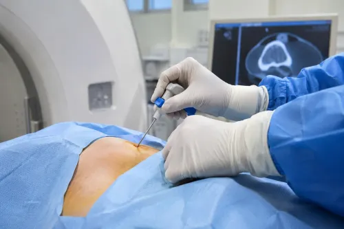
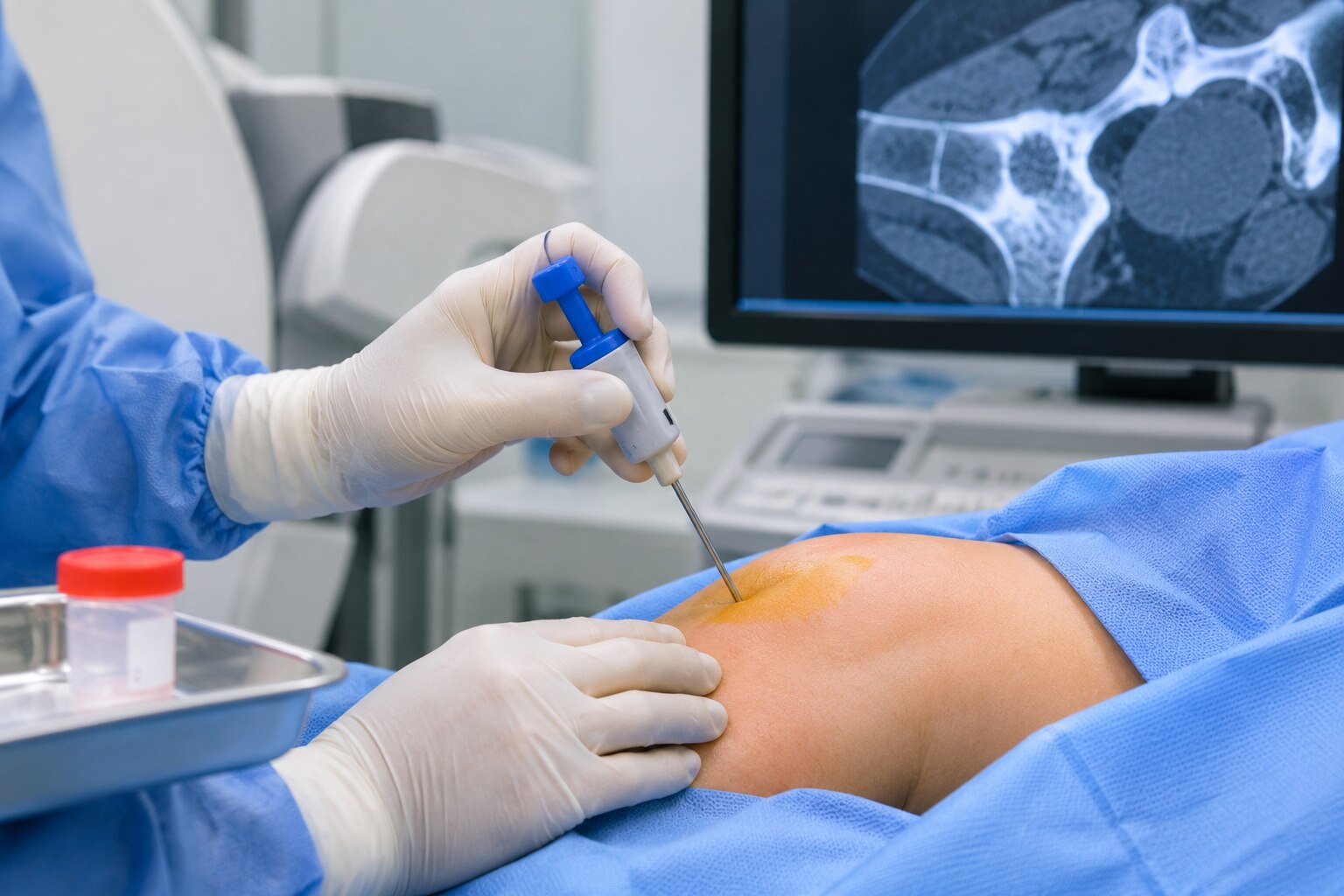
3. Image guidance directs the needle into the target. Several core passes may be needed. The operator checks each sample for adequate tissue before removing the needle.
4. The referral should state suspected myeloma, possible infection and the exact clinical question. Formalin-fixed tissue suits histopathology, but fresh material may be required for flow cytometry, microbiology or molecular tests; these portions are not interchangeable.
5. A dressing covers the site, followed by observation for bleeding, pain or other immediate problems. You receive instructions about keeping it clean, activity, pain relief and when to seek help.
Mild soreness is expected. Report increasing swelling, persistent bleeding, fever, redness, numbness, weakness or breathing difficulty; risks vary by biopsy site.
A benign or nondiagnostic result does not exclude cancer if imaging remains suspicious. The next step may be repeat CT-guided core biopsy, a different target or open biopsy.
How should you choose a biopsy pathway in Mumbai?
There is no universally best needle biopsy option in mumbai. For needle biopsy options for myeloma in mumbai, compare the lesion’s location, size, lytic or sclerotic appearance, soft-tissue extension, suspected diagnosis, bleeding risk, available scans, operator experience and laboratory capacity.
| Pathway | Strength | Main limitation |
|---|---|---|
| CT-guided core biopsy | Reaches deep, pelvic, spinal, sclerotic or complex lesions and obtains tissue for histology | Requires safe planning around vessels, nerves and organs |
| Ultrasound-guided biopsy | Shows superficial lesions or accessible soft-tissue components without radiation | Cannot reach every intraosseous or deep target |
| Marrow biopsy or lesion biopsy | Marrow supports myeloma testing; lesion tissue clarifies an atypical mass or competing diagnosis | They answer different questions and are not interchangeable |
For a needle biopsy for pathological fracture in mumbai, remember that biopsy diagnoses the cause but does not stabilise the bone. Severe pain, deformity, inability to bear weight or suspected spinal instability requires urgent orthopaedic assessment alongside biopsy planning.
Ask these questions before booking:
- Who will select the target and review all scans?
- Has an orthopaedic oncologist checked that the needle track fits the definitive surgical field?
- Which tests need fresh tissue rather than formalin-fixed tissue, including flow cytometry, microbiology or molecular testing?
- What happens if the sample is nondiagnostic?
Interventional radiology, pathology, orthopaedic oncology and oncology should coordinate the plan. Dr Mishil Parikh can help connect biopsy planning with fracture protection, limb-sparing surgery or treatment for a suspected bone tumour, rather than simply choosing the procedure called the best needle biopsy option in mumbai.
Related services
Pathological Fracture A pathological fracture, also known as a pathologic fracture, is a type of bone fracture that occurs as a result of an underlying disease or... View service → |
Core Needle Biopsy Core needle biopsy is a minimally invasive diagnostic procedure used to obtain tissue samples from suspicious areas in the body for further... View service → |
Frequently asked questions
Which tissue sample is needed when myeloma is suspected?
Confirmation usually requires a bone-marrow aspirate and trephine biopsy, which support plasma-cell testing, flow cytometry and genetic studies when indicated.
How does imaging determine the biopsy target and needle route?
MRI, CT or other imaging helps identify the most diagnostic lesion, avoid blood vessels and nerves, and plan a safe needle path.
What changes when a pathological fracture needs urgent care?
The team first assesses stability, pain and the risk of further displacement, then coordinates biopsy timing with fracture protection or urgent surgical treatment.
What happens during a needle biopsy appointment?
After imaging review and consent, the clinician numbs the area, obtains tissue with a needle, and sends labelled samples for the tests requested.
How should you choose a biopsy pathway in Mumbai?
Choose a centre that can coordinate imaging, interventional biopsy, pathology, myeloma testing and orthopaedic oncology review when a fracture or bone lesion is involved.
Related services
Myeloma Myeloma specifically involves abnormal plasma cells that accumulate in the bone marrow and interfere with the production of normal blood cells. |
Image Guided Biopsy Image-guided biopsy is a procedure that uses medical imaging techniques, such as ultrasound, computed tomography (CT), magnetic resonance imaging... View service → |
Keywords