A needle biopsy is most useful when the team can reach the right part of a tumor without crossing important nerves, blood vessels, joints, or tissue compartments. By the end, you will know when imaging adds meaningful safety and diagnostic value, which guidance method fits different tumor locations, and when an open biopsy is the safer plan.
Key takeaways
- Use imaging when the tumor is deep, near vital structures, or difficult to feel.
- Match ultrasound, CT, or MRI guidance to the tumor’s location and visibility.
- Plan the needle track so it can be removed during the definitive operation.
- Ask an orthopaedic oncology team when a needle biopsy is unsafe or inadequate.
When does image guidance improve a complex tumor biopsy?
Image guidance improves a complex tumor biopsy when touch cannot reliably locate the safest path or the most useful tissue. A superficial, soft mass that is easy to feel may be reachable by palpation-guided or freehand placement; a lesion’s position becomes uncertain when it lies:
- Deep or small
- Fixed beneath muscle
- Close to a major nerve or blood vessel
- Embedded in bone
- Within the pelvis
- Surrounded by bowel, joint, or other important anatomy
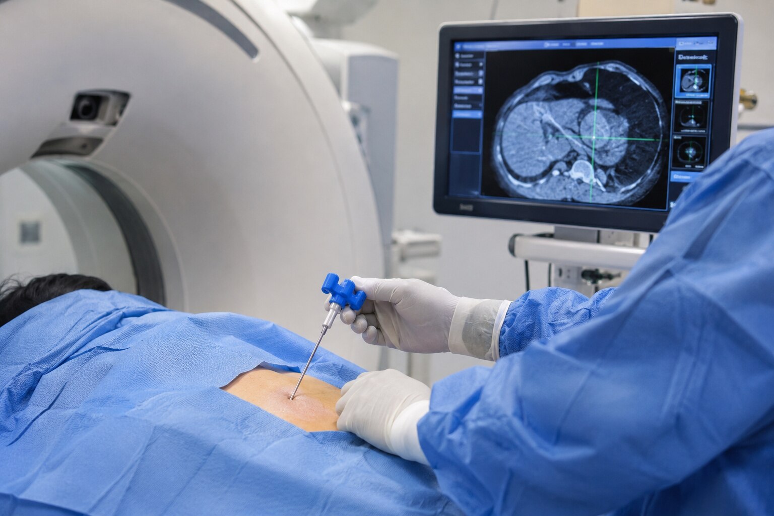
Imaging lets the operator choose a safe entry point and steer the needle toward viable tumor rather than the easiest accessible spot. This matters when a complex tumor contains solid, necrotic, hemorrhagic, cystic, or fibrotic regions; cores taken only from a dead or fluid-filled centre can miss the diagnosis.
For anyone asking when is image guided needle biopsy better for a complex tumor in Mumbai, the city and the equipment name do not decide it. The choice depends on the tumor’s anatomy, imaging appearance, suspected diagnosis, required laboratory tests, and the surgical plan.
Guidance reduces avoidable targeting errors, but it cannot eliminate bleeding, infection, pain, injury to nearby structures, or a nondiagnostic sample. Discuss blood-thinning medicines and bleeding disorders before the procedure.
Which imaging method fits the tumor’s location?
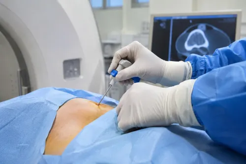
The tumor’s location determines which method gives the operator a safe, visible needle path. MRI or CT usually identifies the target and its viable enhancing regions beforehand; during advancement, the safest guidance is the method that shows the needle and critical structures clearly.
| Method | Best fit | Main advantage | Main limitation |
|---|---|---|---|
| Ultrasound | Superficial soft-tissue masses | Real-time imaging, Doppler vessel mapping, no ionising radiation, and rapid needle adjustment | Bone, air, depth, or larger body size can block the view |
| CT | Deep masses, pelvic or retroperitoneal lesions, and bone tumors | Clearly shows the needle, bone, and surrounding anatomy | Uses ionising radiation and repeated scans are slower and less real-time |
| MRI | Tumors with difficult soft-tissue extent, viable regions, or nerve and vessel relationships | Excellent soft-tissue contrast and assessment of tumor boundaries | Longer procedure time, restricted access, and MRI-compatible equipment requirements |
| Fluoroscopy | Bony landmarks or a joint-related route | Continuous X-ray guidance for positioning along bone or into a joint | Uses radiation and shows soft tissue poorly |
A superficial mass that moves visibly with ultrasound is often easier to sample there than with CT. A deep pelvic lesion hidden behind bowel or bone usually needs CT, while MRI can define which solid region avoids necrosis and important nerves or vessels.
Fluoroscopy suits a bone or joint route, not a mass whose soft-tissue margins are unclear. For anyone comparing image guided needle biopsy in mumbai, location and anatomy matter more than the machine’s name.
Which biopsy type provides the tissue the diagnosis needs?
For a deep or complex tumor biopsy, image-guided core needle biopsy usually provides the most useful balance of diagnostic tissue and limited injury. The choice depends on whether the pathologist needs cells, tissue architecture, or a large specimen.
| Option | What it removes | When it applies |
|---|---|---|
| Fine-needle aspiration | Individual cells | Selected conditions, but often too little architecture to classify sarcoma, assess differentiation or grade, or complete immunohistochemical and molecular tests |
| Core needle biopsy | Cylinders of tissue | Preferred for many deep musculoskeletal tumors; multiple cores can sample different viable regions through a small wound |
| Incisional biopsy | Part of the lesion | Considered when substantially more tissue is needed or properly targeted core samples remain inconclusive |
| Excisional biopsy | The whole lesion | Suitable only when complete removal is appropriate; not an automatic shortcut for suspected sarcoma |
An image-guided core biopsy lets the operator avoid necrotic or hemorrhagic areas and target viable regions seen on imaging. An unplanned excision can spill tumor into surrounding tissue, contaminate additional compartments, and compromise later limb-sparing surgery.
Tell pathology before the procedure if lymphoma, metastasis, infection, or a rare sarcoma is possible. The laboratory may need to divide specimens for flow cytometry, bacterial, fungal, or mycobacterial cultures, immunohistochemistry, and molecular testing; sending everything in formalin can make some of these tests impossible.
Why must the needle route be planned with the final operation?
The needle tract must follow a path the cancer surgeon can remove with the tumor in one specimen. A poorly placed tract that crosses another muscle compartment, a joint, or a neurovascular plane can contaminate those structures and enlarge the eventual resection.
| Route problem | Why it matters | Possible consequence |
|---|---|---|
| Crosses another muscle compartment | Tumor cells may enter tissue outside the main mass | Wider muscle resection |
| Crosses a joint | The tract may involve the joint lining or capsule | More extensive surgery |
| Runs through a neurovascular plane | The route approaches nerves or major vessels | Complex dissection or loss of limb-sparing options |
After reviewing radiographs, MRI, or CT, the team confirms:
- The target region and safest entry path
- The number of core samples required
- Where each specimen will go, such as histopathology, culture, flow cytometry, immunohistochemistry, or molecular testing
- Whether local anaesthesia is sufficient or sedation is needed
Expect questions about anticoagulants, other medicines, previous bleeding, and relevant blood tests. You will receive consent information and fasting instructions if sedation is planned; exact instructions vary with the procedure and your medical condition.
The operator targets viable enhancing tissue rather than a necrotic centre. Multiple cores from radiologically different regions can reduce sampling error, but they cannot guarantee a diagnostic result.
For anyone asking when is image guided needle biopsy better for a complex tumor in Mumbai, route planning with the definitive surgeon matters more than the city or machine.
When is a needle biopsy the wrong choice, and whom should you ask?
An open or incisional biopsy is more appropriate when a needle cannot reach the lesion safely, repeated correctly targeted cores remain nondiagnostic, or the diagnosis needs substantially more tissue architecture. Infection also changes the plan: the team may need separate specimens for histology and bacterial, fungal, or mycobacterial cultures.
An open biopsy can remain safe when the team responsible for definitive tumor surgery plans its incision and tract. An improvised excision can contaminate another muscle compartment, joint, or neurovascular plane and compromise later limb-sparing treatment.
Before choosing a provider for image guided needle biopsy in Mumbai, ask:
- Who reviewed the MRI or CT, and who selected the target and entry route?
- Has the planned orthopaedic-oncology surgeon approved that route?
- Do you need multiple core samples rather than aspiration alone?
- Can the pathology service diagnose sarcoma and lymphoma, assess infection, and arrange cultures, immunohistochemistry, flow cytometry, or molecular testing?
- Will the pathologist receive the imaging and clinical history, with specialist review available for a difficult result?
Someone comparing a guided biopsy specialist in Mumbai should judge this coordination, not equipment alone. Dr Mishil Parikh may be relevant when you need orthopaedic-oncology input for a bone, soft-tissue, or pelvic tumor; the right provider is the team that connects biopsy planning to the definitive operation.
Related services
Image Guided Biopsy Image-guided biopsy is a procedure that uses medical imaging techniques, such as ultrasound, computed tomography (CT), magnetic resonance imaging... View service → |
Core Needle Biopsy Core needle biopsy is a minimally invasive diagnostic procedure used to obtain tissue samples from suspicious areas in the body for further... View service → |
Frequently asked questions
When does image guidance improve a complex tumor biopsy?
It improves biopsy planning when a tumor is deep, poorly defined by touch, close to nerves or blood vessels, located in the pelvis, or positioned where an inaccurate track could affect later surgery.
Which imaging method fits the tumor’s location?
Ultrasound suits many superficial soft-tissue masses, CT provides reliable access to deep or bony lesions, and MRI helps define tumors near nerves, vessels, joints, and other soft tissues.
Which biopsy type provides the tissue the diagnosis needs?
Fine-needle aspiration collects cells, core needle biopsy collects tissue cylinders for architecture and laboratory testing, and an incisional biopsy removes part of a lesion when needle samples cannot answer the diagnostic question.
Why must the needle route be planned with the final operation?
The route should pass through tissue that the surgeon can remove with the tumor. A poorly placed track can contaminate additional compartments and complicate limb-sparing or reconstructive surgery.
When is a needle biopsy the wrong choice, and whom should you ask?
Ask an orthopaedic oncologist before biopsy when imaging suggests a sarcoma, the lesion is near major neurovascular structures, the diagnosis requires more tissue, or the planned route could compromise definitive surgery.
Keywords