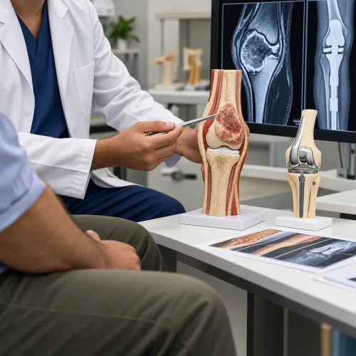

A deep or enlarging lump beside the hip, knee, shoulder, elbow, wrist or ankle needs more than a routine lump removal or an immediately arranged needle test. By the end, you will know which imaging, needle type, tissue handling and biopsy route to discuss, and when a different diagnostic approach is safer.
Key takeaways
- Plan the needle tract so the surgeon can remove it with the tumor.
- Use MRI for soft-tissue tumors and CT for many bone lesions.
- Choose core biopsy when tissue architecture is needed for diagnosis.
- Ask whether the biopsy team has planned the route with the operating surgeon.
Why the biopsy route matters near a joint
A deep tumor beside a joint needs a planned route because the needle tract can affect the operation that follows. The tract should run through tissue the surgeon can remove with the tumor, not across the joint capsule, a major nerve, blood vessel, tendon sheath, growth plate, or several muscle compartments.
A routine biopsy can contaminate otherwise uninvolved tissue planes. That may turn a limited limb-sparing operation into a larger resection, or make reconstruction more difficult. The needle usually targets the tumor outside or within bone; it does not normally puncture the joint.
Imaging comes first. MRI with contrast maps soft-tissue tumors against tendons, muscles, nerves, vessels, and the capsule. Plain radiographs and CT help define bone destruction, cortical involvement, or mineralisation. The operator then chooses a viable solid portion rather than a necrotic or haemorrhagic centre.
Before arranging a needle biopsy for a deep tumor near a joint in Mumbai, confirm that:
- An orthopaedic-oncology team has reviewed the MRI or CT and marked the intended tract.
- The radiologist, sarcoma pathologist, and definitive surgeon will coordinate before the needle enters.
- The planned route avoids uninvolved compartments and remains removable during surgery.
- The team has enough cores for diagnosis, immunohistochemistry, molecular testing, culture, or other studies when indicated.
A technically easy biopsy performed without this plan can produce an inconclusive result or complicate definitive treatment.
Which scan should guide the needle?
Near a joint, the best guidance depends on what the scan shows, not on the joint’s name. MRI with contrast usually maps a soft-tissue mass; CT or fluoroscopy often provides the safer working view for bone lesions.
| Scan | What it shows best | When clinicians use it |
|---|---|---|
| MRI | Tumor extent, viable solid tissue, marrow, capsule, tendons, nerves and vessels | Planning most suspected soft-tissue tumors near the hip, knee, shoulder, elbow, wrist or ankle; the radiologist reviews it before inserting the needle |
| Ultrasound | A moving, superficial target and nearby vessels | A superficial or clearly visible component; it avoids radiation but cannot see through deep bone or large body regions |
| CT | Needle position, cortical bone, mineralisation and deep anatomy | Deep, pelvic, spinal or bone lesions, especially when ultrasound cannot reach the target |
| Fluoroscopy | Real-time X-ray view of bone and instruments | Selected accessible bone lesions or procedures where mineralised landmarks are clear; it gives less soft-tissue detail than CT or MRI |
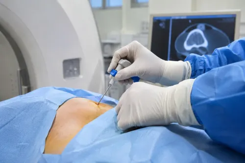
The needle targets the most active, safely reachable solid portion, not a necrotic or haemorrhagic centre. CT guidance is common for a deep lesion, while ultrasound suits a visible superficial component; MRI itself guides fewer biopsies because equipment access is harder, but MRI findings remain essential.
The operator plans a tract that avoids the joint capsule, tendon sheath, major nerve, vessel and uninvolved muscle compartments. In a child, the route also avoids the growth plate when the lesion does not involve it. This planning matters whether you search for soft tissue tumor biopsy in Mumbai or elsewhere.
Fine-needle aspiration versus a core sample
A fine-needle aspiration uses a thin needle to collect loose cells. It can help with selected metastatic deposits, cystic lesions or suspected lymphoma, but cells alone may not show the tissue architecture needed to classify sarcoma or separate it from a benign mimic.
| Sample | What it provides | When it is used |
|---|---|---|
| Fine-needle aspiration | Individual cells for cytology | Selected lesions, recurrence or suspected metastasis; flow cytometry may help when lymphoma is suspected |
| Core needle sample | Cylinders of tissue showing architecture | Deep musculoskeletal masses when sarcoma, lymphoma or an unusual benign tumor is possible |
| Multiple core samples | Material for separate laboratory tests | When diagnosis may require immunohistochemistry, culture, flow cytometry or molecular testing |
The radiologist and pathology laboratory should agree on tissue handling before the procedure. Place representative cores in formalin for histology and immunohistochemistry; send a sterile unfixed portion for culture when infection is a concern, and keep fresh tissue in the laboratory’s specified medium for flow cytometry.
Reserve suitable tissue for cytogenetic or molecular testing rather than putting every fragment into formalin.
A bone tumor needle biopsy needs extra planning. Sclerotic, heavily mineralised or necrotic areas can produce scant, misleading tissue, so the team targets viable solid tumor and may take several cores. Decalcification can damage DNA, RNA and tissue antigens; ask whether a non-decalcified component or validated decalcification method is available.
A negative or scant result does not exclude malignancy when imaging remains suspicious. Include the exact site, bone or soft-tissue origin, imaging findings, cancer history and suspected diagnosis on the pathology request.
How the team avoids a nondiagnostic or unsafe result
A negative core does not overrule suspicious MRI or CT findings. The radiologist targets the most viable, solid and safely reachable part of the lesion, avoiding a necrotic or haemorrhagic centre. Mineralised or sclerotic bone can produce scant tissue, so several cores may be taken.
For a bone lesion, the team may preserve a non-decalcified soft-tissue component because acid decalcification can damage DNA, RNA and tumour markers needed for immunohistochemistry or molecular testing. A small calcified fragment cannot support every test.
Before proceeding, the team checks:
- Anticoagulant and antiplatelet medicines
- Previous abnormal bleeding
- Platelet count and clotting tests when indicated
Correct an avoidable bleeding risk before biopsy. A lesion beside a major vessel, nerve, tendon, joint capsule or a child’s growth plate requires a route that avoids those structures and follows a line the definitive surgeon can remove with the tumour.
Passing through several muscle compartments or across the joint can contaminate tissue planes and enlarge the later operation.
Delay the procedure if bleeding risk is not controlled or no safe target exists. If the sample is inadequate, correlate the pathology with imaging and repeat an image-guided core biopsy from a better target.
Choose an open biopsy when repeat needle sampling cannot safely obtain viable tissue or remains nondiagnostic; the definitive surgical team must plan it. Never substitute an unplanned excision for diagnosis when sarcoma is possible.
Choosing a biopsy team in Mumbai
Before arranging a needle biopsy near joint in Mumbai, verify that the radiologist, orthopaedic oncologist, surgeon and sarcoma-experienced pathologist can coordinate on one plan. The biopsy tract should follow a route the definitive surgeon can remove with the tumour; a poorly placed tract can contaminate muscle compartments and complicate limb-sparing surgery.
Ask the facility these questions:
- Has the radiologist reviewed the complete MRI or CT before choosing the target and needle path?
- Will the planned tract avoid the joint capsule, tendon sheath, major nerve, blood vessel and uninvolved muscle compartments?
- Does the pathology laboratory diagnose bone and soft-tissue sarcomas and perform immunohistochemistry?
- Can the team reserve tissue for culture, flow cytometry or molecular testing when the imaging or pathology requires it?
- Who will review a nondiagnostic result and decide between repeat core biopsy and a planned open biopsy?
| Arrangement | What to verify | Why it matters |
|---|---|---|
| Radiology-only facility | Written tract plan and confirmed receiving pathology laboratory | A technically successful sample can still leave the surgical team with a contaminated route or inadequate diagnosis |
| Multidisciplinary sarcoma service | Imaging, biopsy, pathology and definitive surgery coordinated before the procedure | The team can target viable tumour and preserve tissue for necessary tests |
| Separate surgeon and laboratory | Named communication between both teams and a plan for a nondiagnostic result | Discordant imaging and pathology should trigger review, not unplanned tumour removal |
Dr Mishil Parikh is relevant to this decision because the definitive surgeon should help approve the biopsy route before the needle enters tissue. Do not accept immediate unplanned excision as a substitute for diagnosis when sarcoma is possible.
Related services
Core Needle Biopsy Core needle biopsy is a minimally invasive diagnostic procedure used to obtain tissue samples from suspicious areas in the body for further... View service → |
Image Guided Biopsy Image-guided biopsy is a procedure that uses medical imaging techniques, such as ultrasound, computed tomography (CT), magnetic resonance imaging... View service → |
Frequently asked questions
Why does the biopsy route matter near a joint?
The needle tract should pass through tissue that can be removed with the tumor, without crossing the joint capsule, major nerves, blood vessels, tendon sheaths or multiple muscle compartments.
Which scan should guide a needle biopsy near a joint?
MRI usually maps soft-tissue tumors and their relationship to the joint, tendons and neurovascular structures. CT provides precise guidance for many bone lesions and areas that MRI does not show clearly.
What is the difference between fine-needle aspiration and a core biopsy?
Fine-needle aspiration collects cells for cytology. A core biopsy removes small tissue cylinders, preserving architecture and often providing more information for classifying a bone or soft-tissue tumor.
How can the team reduce the risk of a nondiagnostic or unsafe biopsy?
Review the full imaging set, select a direct removable tract, avoid critical structures, target viable solid tumor rather than necrotic tissue, and coordinate the plan with the surgeon who may remove the tumor.
How should you choose a biopsy team in Mumbai?
Ask whether the radiologist and musculoskeletal oncology surgeon reviewed the imaging together, which scan will guide the procedure, whether core tissue is planned, and how the tract will be removed during definitive surgery.
Keywords