When a soft-tissue lump could be a sarcoma, removing it immediately can make the next operation larger and less effective. You will learn when needle biopsy comes first, how the biopsy route affects surgery, and which questions to ask before tissue is sampled or a lump is removed.
Key takeaways
- Needle biopsy usually comes before definitive sarcoma surgery.
- Do not remove a suspicious deep or enlarging lump without specialist planning.
- The biopsy route must align with the later surgical incision.
- Ask for coordinated imaging, biopsy, pathology, and surgical review in Mumbai.
When sarcoma is suspected, biopsy usually comes before surgery
Needle biopsy usually comes before definitive surgery when a soft-tissue sarcoma is possible. If you are searching “soft tissue sarcoma surgery or needle biopsy which comes first in Mumbai,” the answer is planned assessment first, not immediate lump removal.
| Pathway | What happens | When it fits |
|---|---|---|
| Suspected sarcoma | Examination, MRI or other appropriate imaging, image-guided core needle biopsy, pathology review, local and metastatic staging, then wide excision | Growing, deep, fixed, recurrent, painful, or suspicious mass |
| Small benign-appearing lump | Direct removal may provide the diagnosis | Very small, superficial, freely mobile mass when sarcoma is not suspected |
| Urgent or well-characterised case | Treatment may follow a different sequence | Clinical urgency or an established diagnosis makes the usual pathway unsuitable |
The usual sequence is:
- Examine the mass and obtain MRI when it is deep or near important structures; ultrasound or CT may be appropriate in other settings.
- Plan the biopsy route with the surgeon and radiologist, then take image-guided core tissue.
- Have a sarcoma-experienced pathologist review the specimen for diagnosis, subtype, and grade.
- Complete local staging and assess for spread, including chest imaging when indicated.
- Plan definitive wide excision, removing the tumour with an appropriate cuff of normal tissue while preserving function where possible.
An urgent case or an already well-characterised tumour can require another pathway. That does not make unplanned excision routine for a suspicious mass.
Why removing a suspicious lump first can create a bigger operation
An unplanned excision can turn a manageable sarcoma operation into a wider, more complex procedure. The incision and instruments can spread tumour cells into surrounding tissue planes, while the lump’s margin may be cut through and left tumour-positive.
The first operation can also cross muscle compartments or make the original tumour bed difficult to identify after the lump has been removed. The surgeon treating the sarcoma may then need to remove the scar, the entire biopsy or incision tract, and contaminated tissue around the original site.
That re-excision can sacrifice more muscle, skin, or other structures than planned surgery would have required.
Arrange sarcoma-focused assessment before removal if the mass is:
- Growing, especially rapidly
- About 5 cm or larger
- Deep to the fascia or within muscle
- Fixed to surrounding tissue
- Recurrent after earlier removal
- Associated with unexplained pain
A rapidly enlarging or deep mass is not a routine lump, even when it feels soft, appears harmless, or causes little discomfort. Imaging and a planned biopsy allow the team to map the tumour and place the biopsy route where it can be removed with the tumour.
Removing it first can compromise clear margins and limb-sparing treatment.
Which biopsy is appropriate, and what happens during needle sampling
Core needle biopsy is usually the best first test for a suspected sarcoma because it preserves tissue architecture. That structure helps the pathologist determine subtype and grade, perform immunohistochemistry, and order selected molecular tests without creating the larger wound of an open biopsy.
| Option | What it provides | When it applies |
|---|---|---|
| Core needle biopsy | Several tissue cylinders for architecture, grade, immunohistochemistry, and selected molecular tests | Preferred for most suspected soft-tissue sarcomas |
| Fine-needle aspiration | Loose cells, often too few to establish subtype or grade | Selected known lesions or suspected recurrence |
| Incisional biopsy | A larger open tissue sample | When core samples remain inconclusive or the lesion needs planned open sampling |
| Excisional biopsy | Removal of the entire mass | Only for a small, superficial, readily removable lump when sarcoma is not suspected |
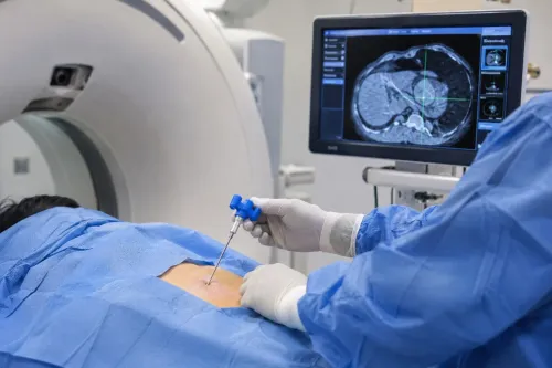
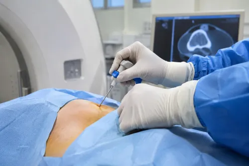
During a needle biopsy for sarcoma, the team reviews MRI or other imaging, cleans the skin, and injects local anaesthetic. Image guidance directs the needle into the target; several passes collect tissue cores, followed by a dressing. You usually return home or remain briefly for observation.
Expect bruising and soreness for a few days. A nondiagnostic sample does not prove the mass is benign: a large or mixed tumour can be missed by sampling. The next step may be repeat image-guided core sampling or a planned incisional biopsy.
When arranging a soft tissue sarcoma biopsy in Mumbai, ask whether a sarcoma-experienced pathologist will review the cores and whether the biopsy route was planned with the treating surgeon.
The biopsy route and imaging plan can determine the later surgical field
The needle tract matters because it is treated as potentially contaminated with tumour cells. Keep it short, straight, and longitudinal on a limb, with the planned operation able to remove it en bloc with the tumour.
An improvised route across an uninvolved muscle compartment or joint can contaminate tissue that would otherwise remain outside the surgical field.
Imaging determines the safest route:
- Ultrasound can guide a visible, accessible superficial lesion.
- CT helps target deep masses or areas that ultrasound cannot show clearly.
- MRI is central for an extremity biopsy because it maps the mass against muscle compartments, fascia, bone, vessels, and nerves.
The choice depends on depth, visibility, and nearby structures—not patient preference alone. A biopsy planned without reviewing the MRI can create avoidable difficulty during limb-sparing surgery.
After sampling, a sarcoma-experienced pathologist reviews the tissue. The report assesses subtype and grade, with selected immunohistochemical or molecular tests when the diagnosis requires them. Chest imaging is added when indicated, particularly when tumour features raise concern for lung metastases, and the team completes multidisciplinary planning before surgery.
A wide excision removes the tumour with an appropriate cuff of normal tissue while preserving useful limb function where possible. The final field depends partly on compartment involvement, neurovascular proximity, tumour biology, and whether the biopsy tract can be removed with the specimen.
How to organise the right biopsy-to-surgery pathway in Mumbai
Before arranging sarcoma surgery in Mumbai, confirm that biopsy and definitive treatment are planned as one pathway, not as separate appointments.
Ask these questions:
- Who will plan the biopsy tract, and has the surgeon approving definitive surgery approved its route?
- Will imaging guide a core-tissue biopsy rather than an improvised or purely fine-needle sample?
- Can the laboratory perform histology, grading, immunohistochemistry, and selected molecular tests?
- Will a pathologist experienced in sarcoma review the specimen?
- Have the surgeon and radiologist examined the MRI together, including the planned needle path?
The key question is not simply where the biopsy is performed. The route must be short, usually longitudinal on a limb, and positioned so the tract can be removed with the tumour; an unapproved route can contaminate tissue planes and enlarge later surgery.
Dr Mishil Parikh can join this specialist discussion when a patient in Mumbai needs coordinated orthopaedic oncology assessment.
Surgery, radiotherapy, and reconstruction are planned together:
| Option | Main advantage | Main trade-off |
|---|---|---|
| Preoperative radiotherapy | Smaller treatment field | Higher wound-complication risk |
| Postoperative radiotherapy | Treatment follows surgical findings | Larger field and different late tissue effects |
A sarcoma-experienced team balances these choices with function and reconstruction. When sarcoma is suspected, arrange the planned biopsy before definitive removal.
Related services
Soft Tissue Tumor A soft tissue tumor refers to an abnormal growth of cells that occurs in the soft tissues of the body. View service → |
Core Needle Biopsy Core needle biopsy is a minimally invasive diagnostic procedure used to obtain tissue samples from suspicious areas in the body for further... View service → |
Frequently asked questions
Does needle biopsy come before soft tissue sarcoma surgery?
Yes. When a soft-tissue sarcoma is possible, planned imaging and needle biopsy usually come before definitive surgery.
Why can removing a suspicious lump first create a bigger operation?
An unplanned removal can contaminate surrounding tissue and force a wider second operation to clear the biopsy site.
Which biopsy is used for suspected soft tissue sarcoma?
A core needle biopsy is commonly used because it provides tissue for diagnosis and tumour grading while limiting disruption to surrounding tissue.
Why does the biopsy route matter for later sarcoma surgery?
The biopsy tract can contain tumour cells, so surgeons plan its position for removal during definitive surgery.
How should you organise sarcoma biopsy and surgery in Mumbai?
Arrange imaging, a planned needle biopsy, pathology review, and surgical consultation through a team experienced in musculoskeletal tumours.
Related services
Image Guided Biopsy Image-guided biopsy is a procedure that uses medical imaging techniques, such as ultrasound, computed tomography (CT), magnetic resonance imaging... View service → |
Limb Salvage Surgery also known as limb-sparing surgery, is a procedure performed to remove a tumor or manage a severe injury in a limb while preserving the limb itself. View service → |
3D Printed & Computer Navigation Assisted Cancer Surgery 3D Printed & Computer Navigation Assisted Cancer Surgery 3D printing and computer navigation technologies have revolutionized the field of cancer surgery, particularly in complex cases that require precise... View service → |
Keywords