Chondrosarcoma is a type of bone cancer characterized by malignant cells that produce cartilage-like tissue. Unlike some other primary bone cancers that are more frequently seen in children and teenagers, conventional chondrosarcoma is more commonly diagnosed in adults.
Its behaviour can vary considerably. Some tumors grow relatively slowly, while higher-grade forms can behave more aggressively. Accurate diagnosis therefore plays an important role in determining treatment.
Where Can Chondrosarcoma Occur?
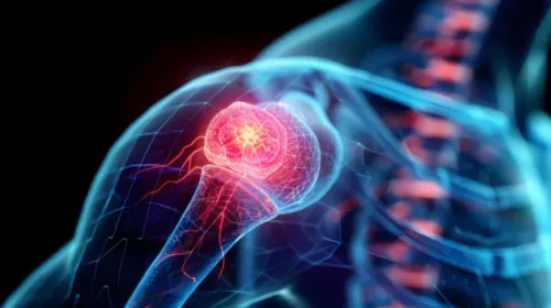
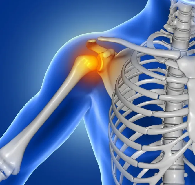
Chondrosarcoma can affect several bones, including the pelvis, thigh bone, upper arm and shoulder region.
Because some tumors develop slowly, symptoms may be present for a significant period before diagnosis.
Patients may notice
- Persistent localized pain
- Pain that gradually increases
- Swelling
- A growing mass
- Reduced joint movement
- Pressure-related symptoms depending on location
A cartilage lesion detected on imaging does not automatically mean chondrosarcoma. Several benign cartilage tumors can also occur, making expert interpretation important.
How Is a Suspicious Cartilage Tumor Assessed?
Assessment begins with clinical history and imaging. X-rays can show characteristic changes within bone, while MRI helps evaluate the tumor's extent and its relationship with nearby tissues.
CT imaging may provide additional information in selected cases.
Doctors consider multiple features rather than relying on a single scan finding. The patient's age, symptoms, lesion location, imaging characteristics and changes over time can all contribute to assessment.
When tissue confirmation is required, biopsy must be carefully planned.
Why Tumor Grade Matters
Chondrosarcoma is not one uniform disease. Pathologists assess characteristics of the tumor to help determine its type and grade.
Tumor grade provides information about biological behaviour. Higher-grade tumors generally have a greater potential for aggressive local growth and spread.
This information influences surgical planning and follow-up.
There are also different subtypes of chondrosarcoma, some of which have distinct behaviour and treatment considerations. Accurate pathology is therefore central to the care plan.
What Role Does Surgery Play?
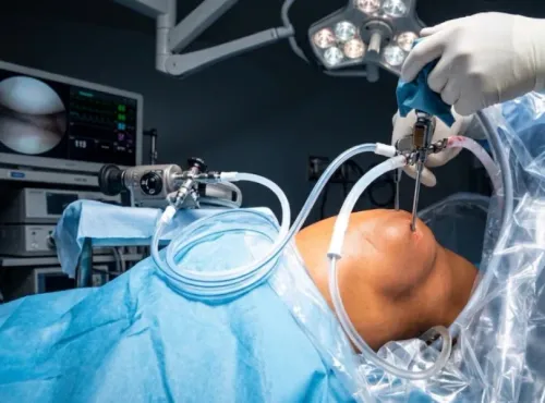
For many conventional chondrosarcomas, surgery is a central part of treatment. The goal is to remove the tumor with an appropriate margin while preserving as much useful function as safely possible.
The operation required depends heavily on where the tumor is located.
A tumor in the pelvis presents different reconstructive challenges from one near the knee or shoulder. Surgeons must consider tumor clearance alongside nerves, blood vessels, joints, muscles and future mobility.
When chondrosarcoma occurs in an extremity, limb-salvage techniques may be considered when appropriate.
Why Is Treatment Planning Individualised?
Two people diagnosed with chondrosarcoma may need very different operations. Factors affecting treatment include
- Histological subtype
- Tumor grade
- Tumor size
- Anatomical location
- Involvement of surrounding structures
- Presence of disease elsewhere
- Patient's general health
- Expected function following surgery
This is why a treatment recommendation cannot be based on the word chondrosarcoma alone.
Living in Mumbai and Planning Treatment
Patients in Mumbai have access to imaging, pathology, medical oncology, radiation oncology, surgical oncology and rehabilitation services across the city. For musculoskeletal tumors, coordination among these disciplines is particularly valuable.
Dr Mishil Parikh assesses patients with suspected and confirmed bone cancers, including chondrosarcoma, with attention to surgical planning and preservation of function when feasible.
Before proceeding with treatment, patients should understand exactly what pathology has shown, the extent of the tumor, the purpose of the proposed operation and the likely functional impact.
Chondrosarcoma requires careful evaluation because its behaviour ranges across different grades and subtypes. A diagnosis based on reliable pathology and appropriate imaging provides the foundation for choosing a treatment plan suited to the individual tumor.
Keywords