An MRI can show where a bone lesion is and how it behaves, but it cannot identify every tumour by appearance alone. By understanding what the tissue sample can establish, which laboratory tests may be added, and how the biopsy route affects later surgery, you can ask more useful questions before treatment begins.
Key takeaways
- MRI maps the mass; tissue identifies its tumour type.
- Core biopsy can guide the surgical plan before the operation.
- A planned biopsy route helps prevent contamination of healthy tissue.
- Ask who will perform, review and coordinate your biopsy in Mumbai.
What can tissue confirm that an MRI cannot?
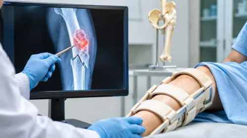
An MRI can show where a bone mass is, how large it appears and whether it involves nearby muscle, nerves or blood vessels. It cannot reliably identify what the mass is. That requires tissue.
A representative needle core can distinguish:
- A benign lesion from a primary malignant bone tumour
- Metastatic carcinoma from a tumour that began in bone
- Lymphoma from sarcoma
- Infection from cancer
- Non-cancerous conditions that mimic cancer on imaging
The sample can establish a tumour family and, when it contains representative tissue, a specific diagnosis such as osteosarcoma, chondrosarcoma, Ewing sarcoma, metastatic carcinoma or lymphoma. Routine microscopy may need support from immunohistochemistry, molecular testing or cytogenetic testing before the pathologist can separate similar-looking diseases.
A result supporting sarcoma is not an isolated label. The pathologist and treating team must compare it with the X-ray, MRI or CT: a small core can miss different areas within a heterogeneous mass, including a higher-grade component.
The scan can also reveal a pattern that makes a biopsy result unexpected and prompts review or repeat sampling.
For readers searching what needle biopsy for confirms before bone tumor surgery in Mumbai, the key point is simple: biopsy identifies the disease in the sampled tissue; imaging shows its location and extent. Both are needed to plan safe treatment.
How does the pathology laboratory identify the tumour type?
The laboratory identifies tumour type by moving from tissue structure to targeted tests. A core biopsy gives the pathologist preserved architecture and cell appearance on routine histology, which can distinguish many benign lesions from malignant tumours and suggest diagnoses such as osteosarcoma or chondrosarcoma.
When microscopy alone cannot separate similar-looking tumours, the pathologist may add:
- Immunohistochemistry, which uses antibodies to detect proteins in tumour cells. These markers help distinguish lymphoma, myeloma, metastatic carcinoma and soft-tissue sarcoma from other small round-cell or spindle-cell tumours.
- Molecular or cytogenetic testing, which detects gene rearrangements, mutations or chromosome changes required for selected diagnoses. Ewing sarcoma, for example, may need testing for a characteristic gene rearrangement; other sarcomas also have defining molecular findings.
- Additional stains or tests when infection, metastatic cancer or an unusual tumour is possible. The laboratory needs suitable tissue, so the specimen may be divided before processing.
A core can also support tumour grading when it contains enough representative, viable tumour. A small sample cannot always capture the highest-grade area of a large or heterogeneous mass, so the biopsy grade may differ from the grade in the resection specimen.
Pathology findings must be read with the X-ray, MRI or CT. If you search for “needle biopsy bone tumor in mumbai,” choose a service where imaging, specimen review and musculoskeletal tumour expertise are coordinated rather than treating microscopy as an isolated answer.
How much can a biopsy establish before the operation?
A biopsy can establish what the sampled lesion is well enough to choose the next treatment, but it cannot map the whole disease. The result may support observation, redirect care towards infection management, prompt systemic treatment, or lead to planned tumour surgery.
The tissue diagnosis can guide the pathway in this order:
- A benign finding that matches the imaging may support observation or scheduled follow-up rather than immediate surgery.
- Organisms, inflammation or infection-related changes shift treatment towards microbiology-guided antibiotics, drainage or other infection management.
- Lymphoma, metastatic cancer and selected sarcomas may require systemic treatment before any operation.
- A resectable primary bone tumour may lead to carefully planned tumour surgery after staging and multidisciplinary review.
Biopsy is not staging. Clinicians still need local MRI to define the lesion’s relationship to bone, muscle, joints and neurovascular structures, plus an assessment for distant disease. For suspected sarcoma, that assessment commonly includes chest imaging; the tumour type determines whether additional scans are needed.
The sample cannot establish the complete tumour size or local extent, prove that all disease has been removed, confirm clear surgical margins, or determine whether limb-sparing surgery is feasible. It also cannot guarantee that every part of a heterogeneous tumour has the same grade.
Those answers depend on MRI, operative findings and examination of the final resection specimen.
Why do the biopsy route and sample quality affect the result?
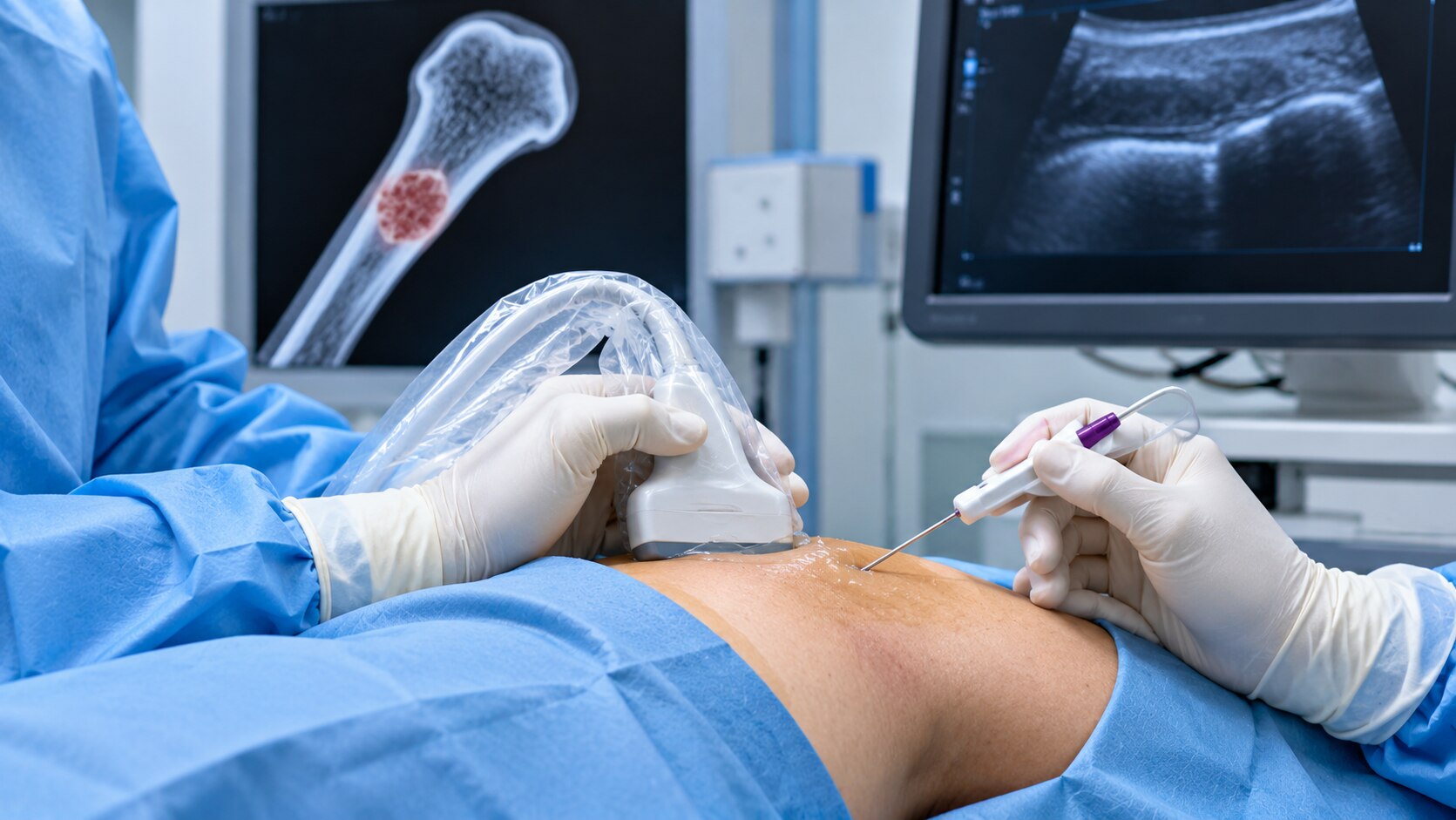
The biopsy should follow adequate X-ray, MRI or CT, because imaging shows the safest route into representative tissue and its relationship to nerves, vessels and planned margins. An orthopaedic oncologist, image-guided radiologist and bone-and-soft-tissue pathologist should agree on the target, route, number of cores and specimen handling before the needle enters.
| Method | Strength | Main limitation |
|---|---|---|
| Core needle biopsy | Removes tissue cylinders for architecture, grading and ancillary tests | Can miss a heterogeneous tumour |
| Fine-needle aspiration | Quickly samples cells or fluid | Often lacks architecture needed for sarcoma classification |
| Incisional or open biopsy | Provides a larger tissue sample | Leaves a surgical wound that must be placed along the future resection |
| Excisional biopsy | Removes the whole lesion | A poor first choice for many bone masses because it can contaminate compartments and compromise planned limb-sparing surgery |
Target viable enhancing tissue, not necrotic, cystic or haemorrhagic areas. Collect enough cores for routine histology, immunohistochemistry and molecular testing; send separate material for bacterial, fungal or mycobacterial cultures and special stains when infection is possible.
For a core needle biopsy before surgery in Mumbai, ask whether the tract will be documented and removed en bloc with the tumour. A transverse incision or route crossing several compartments can force wider resection.
A non-diagnostic result does not prove benign disease. Sampling necrosis, haemorrhage or reactive tissue can prompt multidisciplinary review, repeat image-guided core biopsy or a planned open biopsy when imaging remains concerning.
What should you ask a Mumbai team before scheduling the biopsy?
1. Ask whether an orthopaedic oncologist has reviewed the X-ray, MRI or CT before anyone samples the lesion. The team should explain the safest target, avoiding necrotic or cystic areas, and how the biopsy result will affect treatment.
2. Ask who will plan and document the biopsy tract. It must be marked on the imaging and positioned so the surgeon can remove it with the tumour; an unplanned route through multiple tissue compartments can complicate limb-sparing surgery.
3. Confirm that care includes an orthopaedic oncologist, an image-guided interventional radiologist and a pathologist experienced in bone-and-soft-tissue tumours. Someone searching for a bone tumor biopsy specialist in Mumbai should compare this coordination, not just the person holding the needle.
An integrated service such as Dr Mishil Parikh’s connects biopsy interpretation with subsequent musculoskeletal tumour surgery.
4. Ask how many cores the team plans to collect and how the specimen will be divided for histology, immunohistochemistry, molecular testing or microbiology. If infection is possible, confirm that tissue will reach the laboratory for cultures and special stains rather than going entirely to histology.
5. Ask whether the procedure uses local anaesthetic alone or local anaesthetic with sedation, and what recovery restrictions apply. Discuss pain, bleeding, infection, injury to nearby nerves or vessels, and a non-diagnostic sample; the last problem can require a repeat core biopsy or planned open biopsy.
Related services
Core Needle Biopsy Core needle biopsy is a minimally invasive diagnostic procedure used to obtain tissue samples from suspicious areas in the body for further... View service → |
Image Guided Biopsy Image-guided biopsy is a procedure that uses medical imaging techniques, such as ultrasound, computed tomography (CT), magnetic resonance imaging... View service → |
Frequently asked questions
What can tissue confirm that an MRI cannot?
MRI shows the mass’s location, size and relationship with nearby structures. Tissue can identify whether it is benign or malignant and help determine the tumour type.
How does the pathology laboratory identify the tumour type?
The laboratory examines biopsy tissue under a microscope and uses additional tests when needed to classify the tumour.
How much can a biopsy establish before the operation?
A biopsy can provide a diagnosis and information that helps the surgical team plan treatment. It cannot always establish the tumour’s full extent or replace imaging.
Why do the biopsy route and sample quality affect the result?
The needle route must fit the planned operation because the biopsy track can contain tumour cells. An inadequate or poorly preserved sample can limit diagnosis and delay treatment.
What should you ask a Mumbai team before scheduling the biopsy?
Ask who will perform the biopsy, which imaging will guide it, how the route fits the planned surgery, who will examine the tissue and when you will receive the report.
Keywords