A fracture through weakened bone needs more than a plate, nail or plaster: doctors must identify why the bone failed before choosing durable treatment. You will learn which specialist should lead care, when to go to an emergency department, what tests and biopsy planning involve, and how to select a coordinated Mumbai team.
Key takeaways
- Choose an orthopaedic oncologist to coordinate tumour and fracture treatment.
- Seek emergency care for severe pain, deformity, numbness, or a cold limb.
- Do not arrange fracture fixation before biopsy planning by the tumour team.
- Bring imaging discs, reports, prescriptions, and prior biopsy results to Mumbai consultation.
Which specialist should lead treatment for a tumor-related fracture?
The answer to “which specialist treats a tumor related pathological fracture in mumbai” is an orthopaedic oncologist, also called a musculoskeletal oncologist or bone-and-soft-tissue tumour surgeon. This specialist coordinates fracture care with diagnosis, biopsy planning, tumour removal, reconstruction and rehabilitation.
| Specialist | Primary responsibility | Limitation |
|---|---|---|
| Orthopaedic oncologist | Leads the tumour-related fracture plan and definitive surgery | Needs coordinated oncology and pathology input |
| General orthopaedic surgeon | Provides fracture assessment or temporary stabilisation | Should not usually plan tumour surgery alone |
| Trauma surgeon | Manages urgent injuries, immobilisation and life-threatening problems | Does not replace tumour-specific diagnosis and reconstruction |
| Medical oncologist | Treats systemic disease such as metastatic cancer, lymphoma or chemotherapy-sensitive tumours | Cannot mechanically stabilise an unstable bone |
| Radiation oncologist | Uses radiotherapy for pain and local tumour control | Radiotherapy does not replace fixation when the bone is mechanically unstable |
| Interventional radiologist | Performs image-guided biopsy or procedures such as tumour embolisation | Must follow the definitive surgical team’s plan |
Choose a centre with orthopaedic oncology, musculoskeletal radiology, tumour pathology, medical oncology and radiation oncology access. Ask whether the case goes to a multidisciplinary tumour board, not merely whether the doctor treats fractures.
Do not arrange an independent biopsy before imaging and orthopaedic oncology review. An incorrectly placed biopsy track can contaminate tissue planes and enlarge the eventual operation. A trauma or general orthopaedic surgeon can protect the limb urgently, but the orthopaedic oncologist should lead the definitive pathway.
When is a suspected tumor-related fracture an emergency?
Treat a suspected tumor-related fracture as an emergency when severe pain follows minor trauma, the limb looks deformed, swelling is rapidly increasing, or you cannot bear weight. Numbness, weakness, a cold or pale limb, absent pulses, fever, or new back pain with leg weakness or bladder and bowel problems also require immediate assessment.
- Call emergency services or go to an emergency department, especially if the person cannot be moved safely or has breathing difficulty, heavy bleeding, confusion, or signs of spinal cord compression.
- Keep the injured limb still in the position found. Support it with padded splints or cushions without straightening the deformity.
- Do not allow weight-bearing, forceful manipulation, massage, unsupervised physiotherapy, or attempts to “set” the bone. These actions can worsen bleeding, nerve injury, or displacement.
- During transfer, support the whole limb and check that fingers or toes remain warm and normally coloured. Report increasing pain, numbness, weakness, or loss of pulses immediately.
- Ask the emergency team to arrange appropriate imaging before definitive fixation and to contact an orthopaedic oncologist or musculoskeletal tumour surgeon.
Tumor-related fracture care in Mumbai requires more than rapid plating. Except for life-threatening or urgent neurovascular problems, definitive surgery should wait until imaging and specialist review establish the lesion and the safest reconstruction plan.
How is the cause diagnosed before definitive fixation?
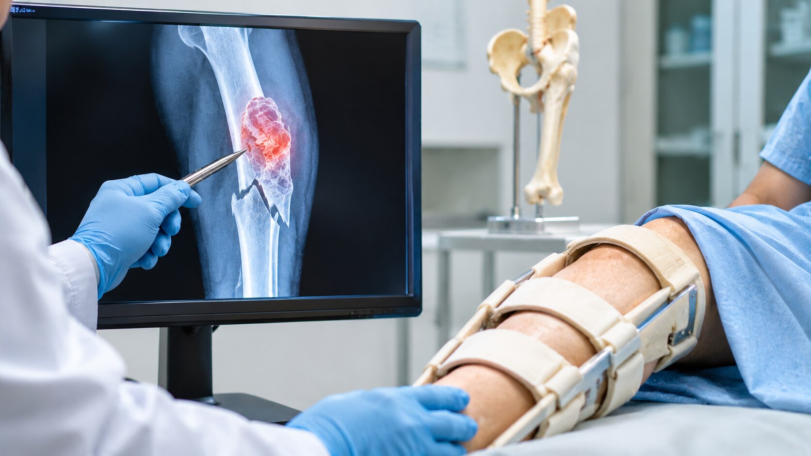
Plain radiographs should cover the entire involved bone, not only the fracture line. This can reveal the lesion’s full length, a second abnormal area and the amount of cortical destruction; missing either can lead to the wrong operation or fixation length.
1. MRI is added when doctors need the lesion’s true marrow extent, soft-tissue spread, blood-vessel or nerve relationships, and the safest surgical corridor. It is especially important when the fracture is near a joint or the mass extends beyond bone.
2. CT shows cortical destruction and complex anatomy more clearly than MRI, helping with fixation and reconstruction planning. It also contributes to staging when doctors suspect spread to the lungs or other bones.
3. Chest imaging, blood tests, CT, a bone scan or FDG-PET/CT are selected when metastasis is possible. The aim is to find the primary cancer and other metastatic sites, not to order every test automatically; the suspected primary tumour and your clinical findings determine the combination.
4. A suspected primary bone tumour should undergo biopsy only after imaging and discussion with the definitive tumour surgery team. The team must place the needle path or incision where it can be removed with the tumour; an unplanned biopsy can contaminate tissue planes and require a larger operation.
Ask whether a bone tumor fracture doctor in Mumbai routinely performs orthopaedic oncology surgery and reviews cases in a multidisciplinary tumour board. That pathway links diagnosis, staging, fixation and tumour treatment instead of treating the fracture alone.
How does the diagnosis change the operation and treatment plan?
The diagnosis determines whether you need tumour removal, durable fracture reconstruction, antibiotics, cancer treatment, or osteoporosis care; routine fixation alone can compromise the next step.
- Metastasis usually calls for immediate pain control and durable fixation, joint replacement, or another reconstruction matched to expected survival and bone destruction. Medical oncology selects systemic treatment, while radiation oncology may add radiotherapy for pain or local control.
- Primary bone sarcoma requires a planned biopsy review followed by wide tumour resection and reconstruction with an endoprosthesis, bone graft, or another method. Chemotherapy and radiotherapy depend on the sarcoma subtype, grade, location, and surgical margins.
- A benign tumour may need curettage, filling with bone graft or cement, and fixation if the bone is unstable. Some lesions need observation instead; radiotherapy and systemic cancer treatment are not routine.
- A bone cyst is treated according to its type and size, with options including aspiration, injection, curettage, grafting, or fixation. The operation changes if imaging or pathology suggests a different lesion.
- Infection requires tissue cultures, surgical drainage or debridement, antibiotics, and stabilisation planned around infection control. Implanting definitive hardware into uncontrolled infection can cause persistent infection and failure.
- Osteoporosis-related fracture needs standard fracture reconstruction or joint replacement plus bone-health assessment and treatment, such as calcium and vitamin D review or prescription osteoporosis therapy. Radiotherapy has no routine role.
A multidisciplinary review should include orthopaedic oncology, musculoskeletal radiology, tumour pathology, medical oncology, and radiation oncology. Ask a pathological fracture specialist in Mumbai whether the proposed operation follows confirmed pathology and tumour-board review, rather than treating the X-ray as an ordinary fracture.
How should you choose and prepare for a Mumbai consultation?
If you are deciding which specialist treats a tumor-related pathological fracture in Mumbai, look for an orthopaedic oncologist, musculoskeletal oncologist, or bone-and-soft-tissue tumour surgeon who routinely operates on tumour-related fractures—not a doctor who only fixes routine breaks.
| Specialist | Role in the decision | Limitation |
|---|---|---|
| Orthopaedic oncologist | Plans biopsy, tumour surgery, reconstruction and fracture stability | Needs oncology and radiology input |
| Trauma or general orthopaedic surgeon | Provides urgent immobilisation or stabilisation | Should not make the definitive tumour plan alone |
| Medical or radiation oncologist | Coordinates drug treatment or radiotherapy | Does not replace mechanical fracture treatment |
| Interventional radiologist | Performs selected image-guided biopsy or procedures | Requires direction from the definitive surgical team |
For reliable tumor related fracture care in mumbai, choose a centre that holds a multidisciplinary tumour board with orthopaedic oncology, musculoskeletal radiology, pathology, medical oncology and radiation oncology. Ask whether a bone-tumour pathologist will review the tissue and whether the reconstruction plan accounts for rehabilitation and later implant failure.
- X-ray and MRI or CT images, with reports and image discs
- Biopsy reports, glass slides or tissue blocks, if already obtained
- Blood-test results, medication list and previous cancer records
- Details of pain, weight-bearing ability, numbness, weakness and the injury
Ask who will plan the biopsy, whether surgery comes before or after radiotherapy, and how the team will protect function. Dr Mishil Parikh is relevant when you want an orthopaedic oncology assessment focused on both the weakened bone and the underlying tumour.
Related services
Pathological Fracture A pathological fracture, also known as a pathologic fracture, is a type of bone fracture that occurs as a result of an underlying disease or... View service → |
Soft Tissue Tumor A soft tissue tumor refers to an abnormal growth of cells that occurs in the soft tissues of the body. View service → |
Frequently asked questions
Which specialist treats a tumor-related pathological fracture in Mumbai?
An orthopaedic oncologist, also called a musculoskeletal oncologist or bone-and-soft-tissue tumour surgeon, should lead care. The team coordinates imaging, biopsy, fracture stabilisation, tumour removal, reconstruction, and rehabilitation.
When is a suspected tumor-related fracture an emergency?
Go to emergency care for uncontrolled pain, an obvious deformity, an open wound, heavy bleeding, new numbness or weakness, a cold or pale hand or foot, or inability to move the limb. Keep the limb still and do not bear weight.
How is a suspected tumor-related fracture diagnosed before definitive fixation?
Doctors review X-rays and usually add MRI or CT to define the lesion and fracture. Blood tests and staging scans can help assess the cause. A planned core-needle biopsy should be placed by the specialist who will perform definitive surgery.
How does the diagnosis change the operation and treatment plan?
A simple fracture may need standard fixation, but a tumour-related fracture requires a plan that accounts for biopsy results, tumour extent, contamination risk, bone quality, and possible removal. Treatment can include tumour surgery, fixation or reconstruction, radiotherapy, systemic therapy, or a combination.
How should you prepare for a Mumbai consultation?
Bring X-ray, CT, and MRI images on discs or secure links, written reports, pathology slides or reports, blood-test results, medication details, and a timeline of pain or injury. Ask who will perform the biopsy, whether fixation should wait, and how limb function will be preserved.
Related services
Sarcoma / Musculoskeletal / Orthopaedic Oncology A bone cyst is a benign, fluid-filled sac that develops within a bone. It is considered a type of benign bone tumor. View service → |
Sarcoma / Musculoskeletal / Orthopaedic Oncology Bone Sarcoma, Benign Bone Tumor Bone sarcoma and benign bone tumors are two distinct types of bone conditions. 1. View service → |
Limb Salvage Surgery also known as limb-sparing surgery, is a procedure performed to remove a tumor or manage a severe injury in a limb while preserving the limb itself. View service → |
Metastatic Spine disease also known as spinal metastasis or metastatic spinal tumor. The symptoms of metastatic spine disease can vary depending on the location and extent of... View service → |
Keywords