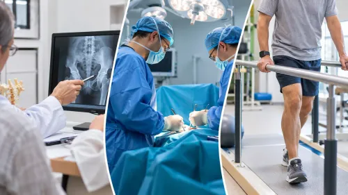
A lump that grows, lies deep, hurts, returns after removal, or measures more than 5 cm needs a planned assessment rather than immediate excision. By the end, you will know how referral, imaging, biopsy, pathology review, treatment, surgery, rehabilitation, and follow-up fit together—and what records and questions to take to a Mumbai sarcoma team.
Key takeaways
- Refer enlarging, deep, painful, recurrent, or over-5-cm lumps before removal.
- Use MRI-guided planning and a properly planned biopsy to confirm sarcoma.
- Compare specialist teams by tumour expertise, imaging, pathology, surgery, and rehabilitation.
- Plan wound care, physiotherapy, surveillance scans, and practical support before surgery.
When a Lump Needs Sarcoma Referral—and What Happens Before It
An enlarging, deep, painful, recurrent, or larger-than-5-cm lump warrants urgent imaging and sarcoma referral in Mumbai. Size alone is not reassuring: a smaller lump can still be malignant. The soft tissue for sarcoma care process in Mumbai should begin with planned assessment, not immediate removal.
Before referral, your first doctor should:
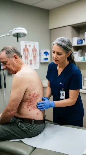
- Examine and record the lump’s size, depth, mobility, tenderness, fixation, skin changes, and rate of growth.
- Arrange ultrasound for a superficial mass; request contrast MRI for a deep, large, or suspicious lesion when appropriate.
- Avoid unplanned excision or “lump removal.” An incision in the wrong place can contaminate tissue planes and make later wide surgery or reconstruction harder.
- Refer the case, images, and clinical notes to a multidisciplinary team before biopsy or treatment. The team should include a musculoskeletal oncologist, sarcoma-experienced radiologist and pathologist, medical and radiation oncologists, and rehabilitation specialists.
| Finding | Action before referral | Why it matters |
|---|---|---|
| Growing or painful lump | Arrange prompt imaging and specialist review | Growth and pain increase concern |
| Deep or fixed mass | Request specialist-directed MRI | The lesion may involve muscle, nerves, or vessels |
| Mass over 5 cm | Make an urgent referral | Size raises the risk of sarcoma |
| Recurrent lump after removal | Send the original records and imaging | Recurrence can reflect an inadequately planned first operation |
Carry original MRI or CT images, not just reports, plus pathology slides or blocks, operative notes, and treatment records. Ask whether the case will receive formal multidisciplinary review before definitive treatment.
From MRI to Biopsy: How the Diagnosis Is Confirmed and Staged
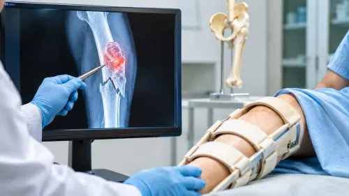
A clear soft tissue sarcoma care process in Mumbai starts with imaging, not immediate removal. The MRI should map the mass before anyone takes tissue, because it shows its relationship to muscle compartments, fascia, nerves, blood vessels, and bone. A lump smaller than 5 cm can still be malignant.
1. Request an MRI of the affected limb or trunk before biopsy. CT may be more suitable for some areas, but the scan must answer where the tumour sits and how far it extends.
2. Plan a core-needle biopsy with the sarcoma team and radiologist. Place the needle track where it can be removed during definitive surgery; an awkward track or unplanned excision can contaminate healthy tissue and widen the operation.
3. Ask pathology to identify the exact subtype and grade, not simply “cancer.” Immunohistochemistry, cytogenetic testing, or molecular testing may be needed. A sarcoma-experienced pathologist should review slides or tissue blocks when the diagnosis is unusual or treatment depends on the subtype.
4. Complete chest staging, usually with a CT chest, because high-grade soft tissue sarcoma commonly spreads to the lungs. Add abdominal, pelvic, bone, or PET imaging only when the tumour type, location, symptoms, or suspected stage supports it.
5. Take the original MRI or CT images, pathology material, reports, and prior treatment records to a multidisciplinary review involving oncology, radiology, pathology, surgery, radiation, medical oncology, and rehabilitation. Treatment should follow that shared review, not an isolated report.
Choosing the Mumbai Team and Comparing Treatment Options
Choose a soft tissue sarcoma specialist in Mumbai by the team behind the clinician, not by the title alone. Look for a musculoskeletal oncologist in Mumbai who routinely manages sarcoma and presents cases with a radiologist, sarcoma-experienced pathologist, medical oncologist, radiation oncologist, reconstructive surgeon, and rehabilitation team.
Ask these questions before accepting a treatment plan:
- How many sarcoma operations does the surgeon perform, and are cases reviewed in a multidisciplinary meeting?
- Who will recheck the MRI images, biopsy slides or blocks, and pathology report?
- Can the team explain the intended margin, reconstruction plan, and effect on limb function?
- Does the plan change if the tumour subtype, grade, or chest staging result changes?
Carry the original MRI or CT images, not only the report, along with pathology material, operative notes, and previous treatment records. Whether you consult Dr Mishil Parikh or another clinician, this review can expose a misclassified tumour or an inadequately planned prior procedure.
Treatment is selected after imaging, biopsy, grade, stage, site, and your health are considered together:
| Option | Main purpose | When the team considers it |
|---|---|---|
| Surgery | Remove the tumour with a negative microscopic margin | Resectable localized disease; preserving function must remain oncologically safe |
| Radiotherapy | Reduce local recurrence risk or treat unresectable disease | Before or after surgery, depending on margins, size, site, and wound risks |
| Systemic treatment | Treat cancer throughout the body | Metastatic, high-risk, or chemotherapy-sensitive subtypes; benefit must outweigh toxicity |
Chemotherapy is not automatic for every localized adult sarcoma. Age, tumour biology, grade, size, and location can change the balance.
Related services
Soft Tissue Tumor A soft tissue tumor refers to an abnormal growth of cells that occurs in the soft tissues of the body. View service → |
Limb Salvage Surgery also known as limb-sparing surgery, is a procedure performed to remove a tumor or manage a severe injury in a limb while preserving the limb itself. View service → |
What Soft Tissue Sarcoma Surgery Involves for Different Body Areas
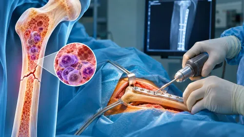
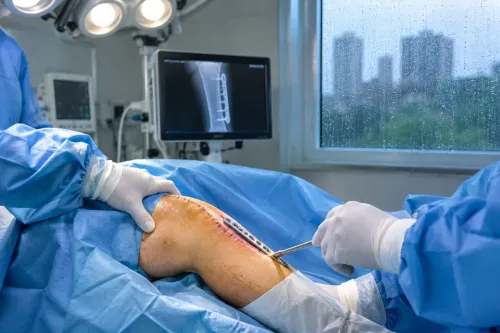
For soft tissue sarcoma surgery in Mumbai, expect an en-bloc wide excision: the tumour and its biopsy tract are removed together, aiming for a negative microscopic margin. There is no universal 1-cm rule; the required margin depends on tumour biology, tissue barriers, anatomy, previous procedures, and radiotherapy.
Reconstruction may use a skin graft, flap, mesh, or tendon repair.
| Body area | Main surgical issue | Site-specific risk |
|---|---|---|
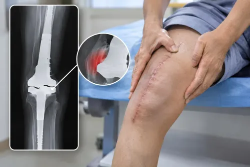
| Arm or leg | Limb-salvage surgery removes tumour while preserving useful nerves, vessels, and muscle | Nerve weakness, vessel injury, stiffness, or reduced strength |
| Pelvis or groin | Resection may involve bone, hip structures, vessels, or nearby organs | Walking difficulty, urinary or bowel problems, and complex reconstruction |
| Trunk or abdominal wall | Wide excision can leave a large soft-tissue defect | Hernia, wound breakdown, or breathing discomfort after chest-wall surgery |
- Limb salvage is chosen when the tumour can be completely removed and the remaining limb will function safely. Amputation enters the discussion when major nerves or vessels are inseparable from the tumour, infection is uncontrolled, or a useful limb cannot be preserved.
- Hospital care depends on the operation. Expect pain control, wound and drain monitoring, blood-clot prevention, and physiotherapy before discharge.
- Ask who will perform reconstruction and how rehabilitation will begin. A sarcoma team should explain the final pathology margin, wound risks, and the signs requiring urgent review.
Recovery, Rehabilitation, Surveillance, and Practical Planning
Recovery begins in hospital, not after the last cancer treatment. The team checks the incision, controls pain, and protects any reconstruction while you regain safe movement.
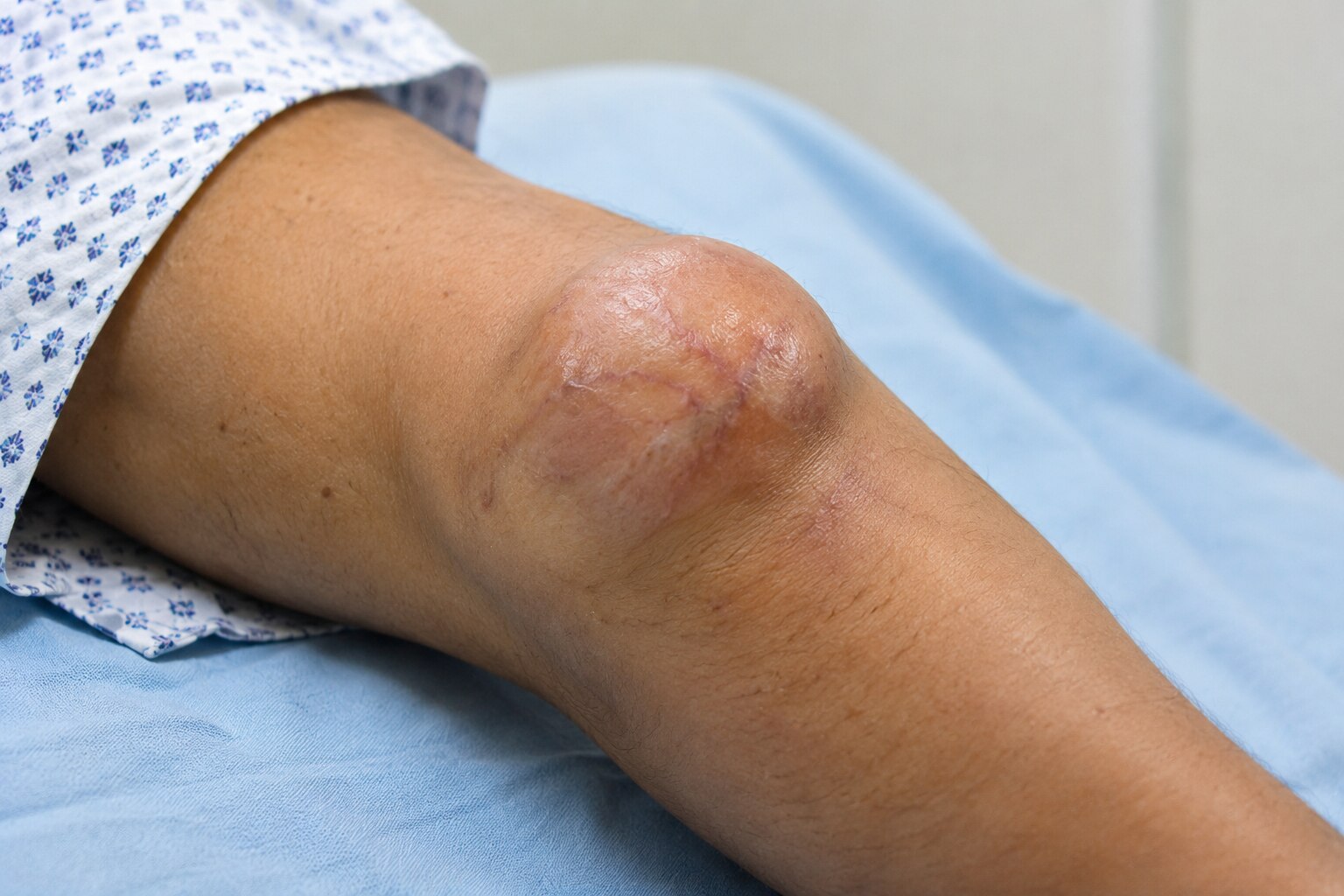
Keep the wound clean and dry as instructed, and do not apply creams or remove dressings without advice. Report increasing redness, drainage, wound separation, fever, or rapidly worsening pain.
Physiotherapy should start at the stage your reconstruction allows. Exercises restore joint movement and strength; gait or limb-use training helps you return safely to daily activities. Ask a soft tissue sarcoma specialist in Mumbai when to begin, which movements are restricted, and how to watch for lymphoedema.
Plan work in stages. Your written plan should cover sitting, standing, lifting, driving, commuting, and follow-up appointments rather than only a return date.
Surveillance depends on the sarcoma subtype, grade, site, treatment, and recurrence risk. Keep every appointment and bring the original scans, reports, and treatment records. Surveillance may include examination, MRI or CT of the treated area, and chest imaging.
Before travelling, confirm that the wound is stable, mobility is safe, medicines are available, and follow-up can continue at your destination. Request a written estimate covering admission, dressings, pathology, scans, physiotherapy, medicines, and travel; ask your insurer about pre-authorisation.
Seek earlier review for:
- A new or enlarging lump
- Persistent or worsening pain
- New swelling, numbness, or limb weakness
- Breathlessness, fever, wound drainage, or wound opening
A practical soft-tissue sarcoma care process in Mumbai includes these checks from discharge through long-term surveillance.
Frequently asked questions
When does a soft-tissue lump need sarcoma referral in Mumbai?
Seek urgent imaging and sarcoma referral for an enlarging, deep, painful, recurrent, or larger-than-5-cm lump. A smaller lump can still be malignant.
How is soft tissue sarcoma diagnosed and staged?
Assessment usually combines clinical examination, MRI, a planned biopsy, pathology review, and staging scans selected for the suspected tumour.
What should you compare when choosing a soft tissue sarcoma specialist in Mumbai?
Compare the team’s experience with sarcoma, access to specialist imaging and pathology, surgical options, multidisciplinary review, rehabilitation, and follow-up planning.
What does recovery after soft tissue sarcoma surgery involve?
Recovery can include wound monitoring, pain control, physiotherapy, mobility or limb-function training, surveillance appointments, and planning for work and daily activities.
Related service
3D Printed & Computer Navigation Assisted Cancer Surgery 3D Printed & Computer Navigation Assisted Cancer Surgery 3D printing and computer navigation technologies have revolutionized the field of cancer surgery, particularly in complex cases that require precise... View service → |
Keywords