An unclear biopsy result does not automatically mean that a tumour is benign, and it does not always mean that another biopsy is the immediate answer. By checking the report wording, comparing the sampled tissue with the scan, and gathering the right records, you can understand whether the next step is review, repeat sampling, surveillance or a planned surgical biopsy.
Key takeaways
- An inconclusive result is not proof that a mass is benign.
- Collect biopsy slides, blocks, report, scans, and clinical history before review.
- Repeat biopsy helps when imaging identifies a safer, more representative target.
- Do not delay specialist assessment when a mass grows or causes new symptoms.
What “inconclusive” means—and why it is not the same as benign
An unclear biopsy result does not mean the mass is benign. It means the specimen did not answer the diagnostic question reliably, either because it contained too little representative tissue or because the cells could not be classified with confidence.
| Report wording | What it means | What it does not prove |
|---|---|---|
| Insufficient tissue | Too little material was obtained for a dependable diagnosis | That the lesion is harmless |
| Nondiagnostic | The sample does not explain the abnormality | That cancer has been excluded |
| Atypical cells | Cells look abnormal but are not definitively malignant | A confirmed cancer diagnosis |
| Suspicious for malignancy | Features raise concern for cancer | The exact tumour type or grade |
| Cannot classify | The tissue is inadequate or too unusual to name confidently | A benign result |
| Necrotic tissue | The needle collected dead tumour or damaged tissue | That all of the lesion is non-cancerous |
| No malignancy identified | No cancer was seen in the submitted material | That cancer is excluded if the sample missed the suspicious area |
“No malignancy identified” is reassuring only when the sample adequately represents the lesion and matches the scan. An enlarging, deep, painful, or larger-than-5-cm mass deserves further review when MRI, CT, or ultrasound remains suspicious. Ask whether the result is radiologically concordant before accepting reassurance.
This matters when reviewing tumor biopsy results unclear in Mumbai or an image guided biopsy unclear result in Mumbai.
Why the needle sample can fail to answer the question
A needle can enter the lesion and still miss the tissue that answers the question. It may collect necrosis, haemorrhage, cyst fluid, scar tissue or reactive inflammation instead of viable tumour. A large mass can also contain several components, including low-grade and high-grade areas, so a technically successful sample can remain nonrepresentative.
Fine-needle aspiration alone may provide scattered cells but not the tissue architecture needed to classify and grade a suspected sarcoma. A scant aspirate cannot reliably exclude sarcoma when the scan remains suspicious.
| Sample or comparison | Why the result stays unclear | What it means |
|---|---|---|
| Necrosis, fluid or haemorrhage | The needle reached a dead, cystic or bleeding area | Viable tumour may remain unsampled |
| Scar tissue or reactive inflammation | Non-tumour tissue surrounded the lesion | The sample may explain the reaction, not the mass |
| Several tissue components | The lesion is heterogeneous | One small core may not represent the most aggressive area |
| Too little tissue or imaging mismatch | There is insufficient material, or pathology conflicts with MRI, CT or ultrasound | The diagnosis needs radiology–pathology review |
Radiology–pathology concordance means the radiologist, pathologist and treating specialist compare the needle path, target, imaging features and pathology before choosing the next step. For a search such as needle biopsy inconclusive tumor in mumbai, ask whether the sampled tissue was viable and representative, not merely whether the procedure was completed.
What to collect before asking for a second opinion
Take the complete case file, not just the radiology summary, when seeking a second opinion for unclear needle biopsy results after image guided biopsy in Mumbai. The summary describes the scan; it cannot show exactly where the needle travelled or whether the tissue came from viable tumour.
- Complete pathology report, including immunohistochemistry results and any addendum.
- Biopsy procedure note, with the imaging method, target, needle path, number of cores or passes, and complications.
- Specimen labels or container details, including the site sampled and specimen type.
- Preserved glass slides or the paraffin tissue block, when available, for expert review.
- Earlier pathology reports, MRI or CT reports, and the actual MRI or CT images on a disc or secure link.
- Earlier scans for comparison, including dates, so the reviewer can assess growth or a changing component.
Ask whether the sample contained viable tumour or only necrosis, haemorrhage, fluid, scar or inflammation. Ask whether the pathology is concordant with the scan, and which exact area was sampled.
Also ask whether the tissue needs expert review, additional immunohistochemistry, fluorescence in-situ hybridisation, cytogenetic testing or sequencing. These tests can classify tumours that look alike under the microscope and may require preserved tissue.
A suspected bone or soft-tissue sarcoma deserves review by a team familiar with biopsy-tract planning. The biopsy route must be considered alongside later surgery because an improperly placed tract can complicate definitive tumour removal.
When a repeat biopsy helps—and how the new target is chosen
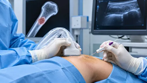
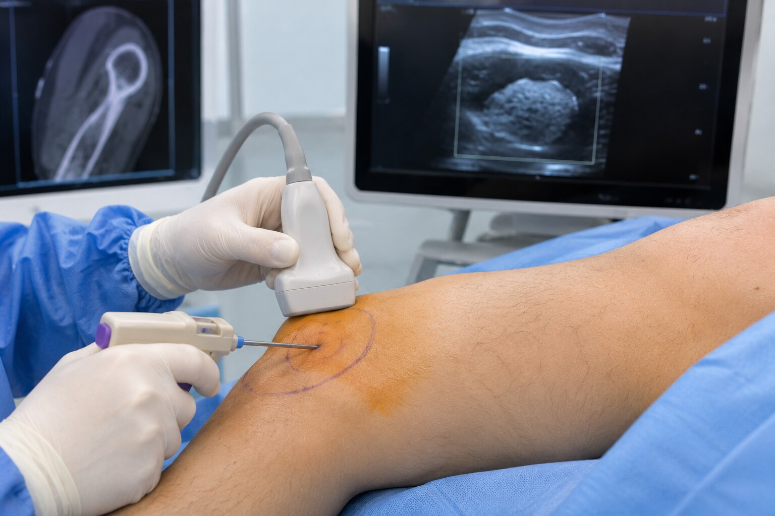
Repeat biopsy after unclear result in Mumbai is appropriate when imaging remains suspicious, the mass grows, the specimen contains mostly necrosis or fluid, or pathology clearly conflicts with MRI, CT or ultrasound. Before repeating the procedure, the radiologist, pathologist and treating specialist should confirm where the needle went and whether it reached viable tumour.
The new target and route are chosen in this order:
- Target the solid enhancing portion on contrast MRI or CT, or the metabolically active portion on PET, rather than a dead, haemorrhagic or cystic centre.
- Map the route around major nerves, blood vessels, bowel and pelvic structures. The needle tract should pass through tissue that can be removed with the tumour during definitive surgery.
- Change the method if the first attempt failed: use a larger-gauge core, a different needle path, or another imaging modality. Do not repeat the identical path into the same nonrepresentative area.
| Option | Strength | Limitation |
|---|---|---|
| New image-guided core | Samples a selected viable region | Still depends on safe access |
| Different needle path or modality | Corrects a targeting problem | Requires new planning and imaging |
| Incisional or open biopsy | Provides more tissue and architecture | Larger procedure with wound and tract concerns |
Dr Mishil Parikh can help coordinate the sampling question with later musculoskeletal tumour surgery, which matters because a poorly placed sarcoma biopsy tract can contaminate tissue planes and complicate limb-sparing or pelvic resection. A technically successful biopsy is not useful if it samples the wrong part of the mass.
Related services
Image Guided Biopsy Image-guided biopsy is a procedure that uses medical imaging techniques, such as ultrasound, computed tomography (CT), magnetic resonance imaging... View service → |
Core Needle Biopsy Core needle biopsy is a minimally invasive diagnostic procedure used to obtain tissue samples from suspicious areas in the body for further... View service → |
When observation is reasonable—and when delay is unsafe
Observation is reasonable when a lesion is stable, the pathology is benign, and the result matches its MRI, CT or ultrasound appearance. Additional imaging or comparison with earlier scans can clarify uncertainty when the first sample was representative and the lesion has not changed.
| Next step | What it means | When it applies |
|---|---|---|
| Scheduled imaging | Watch for growth or new features | Stable lesion with concordant benign pathology |
| Expert pathology review | Re-examine adequate tissue and add tests such as immunohistochemistry, fluorescence in-situ hybridisation or sequencing | Tissue is sufficient but the tumour type remains difficult to classify |
| Repeat core biopsy | Target viable, solid, enhancing tissue | The mass grows, appears aggressive, or the first sample contained necrosis, fluid or scar |
| Planned surgical biopsy | Obtain tissue through a route that can be removed later | Needle sampling remains discordant or cannot safely answer the question |
Delay is unsafe when a mass is enlarging, deep, painful, larger than about 5 cm, radiologically aggressive, or discordant with a benign report. A result labelled benign does not settle an image guided biopsy unclear result in Mumbai if the needle missed the suspicious component.
At the appointment, ask:
- What exact words describe the result?
- Was the sample representative?
- Which area was sampled?
- What is the risk of waiting?
- Who will plan the biopsy tract?
These questions keep decisions about tumor biopsy results unclear in Mumbai focused on concordance, growth and anatomy rather than the word “benign” alone.
Frequently asked questions
Does an inconclusive needle biopsy mean the tumor is benign?
No. It means the specimen did not answer the diagnostic question reliably because it lacked representative tissue or the cells could not be classified confidently.
Why can an image-guided needle biopsy produce an unclear result?
The needle may sample necrosis, blood, scar tissue, inflammation, or a nonrepresentative part of a mixed mass. Small samples can also lack enough tissue for special stains or molecular tests.
What records should you collect before seeking a second opinion?
Request the pathology report, biopsy slides, paraffin tissue blocks, imaging reports, original scans, biopsy procedure note, and a list of symptoms, medicines, and prior treatment.
When does repeating a biopsy help after an unclear result?
A repeat biopsy helps when the first sample was inadequate or imaging shows a different solid, viable, and safer target. The radiologist and treating team choose the target by matching the scan with the diagnostic question.
When is observation reasonable after unclear tumor biopsy results?
Observation is reasonable when imaging and clinical findings support a low-risk process, the lesion is stable, and a specialist has defined follow-up imaging and timing. Prompt reassessment is needed for growth, worsening pain, neurological symptoms, or a changing mass.
Keywords